![]() Figure 1 of
Jablonski, Mol Vis 1999;
5:16.
Figure 1 of
Jablonski, Mol Vis 1999;
5:16.
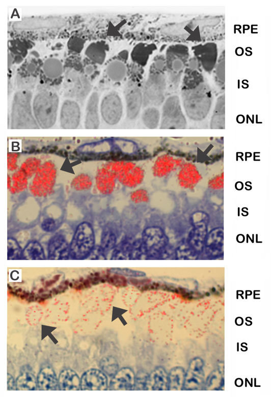
Figure 1. Morphology and immunolocalization patterns of retinas maintained for 3 days in vitro in the presence of an intact retina-RPE complex.
A. In the presence of the RPE, photoreceptor outer segments are composed of an orderly array of stacked discs with no evidence of membranous whorls (arrows). B. Anti-opsin antibody immunolabels heavily in a linear pattern the outer segments of these retinas, indicative of organized discs. C. rds/peripherin is detected at the outer segment disc periphery and in a striated pattern over the outer segment, likely at the incisures (arrows).