![]() Figure 1 of
Uchida, Mol Vis 5:1, 1999.
Figure 1 of
Uchida, Mol Vis 5:1, 1999.
Figure 1. Depolarization with 35 mM extracellular K+ elicits sustained increases of [Ca2+]in of photoreceptor cells.
Cells were treated using Protocol I, as described in Methods. Briefly, following preincubation with Fura-2AM, cells were washed and incubated in 2 ml of BSS. After obtaining 3.5 min (A) or 7 min (B) of baseline data, 0.667 ml of modified BSS containing 129.2 KCl and no NaCl was added to bring the final extracellular K+ concentration to 35 mM. The cells were allowed to remain in this medium for the remainder of the experiment. Fluorescence emission ratios were obtained at 1 min intervals throughout the experiment.
Figure 1A.
Line tracings of [Ca2+]in of six cells in a single microscope field before and for 28 min during depolarization. Pseudocolor animation of the changes in [Ca2+]in can be viewed in Figure 2; the data in the lower left corner of each frame correspond to the emission ratio number and the recording time. All five photoreceptor cells showed sustained increases of [Ca2+]in, while the apparently undifferentiated round cell did not.
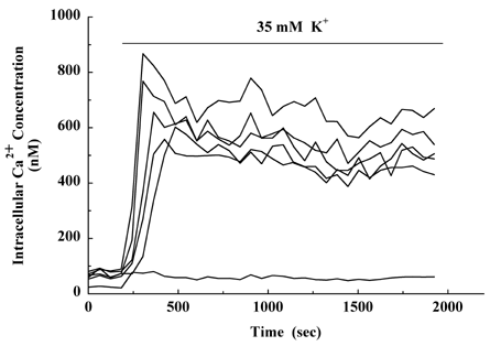
Figure 1B.
Pseudocolor montage of [Ca2+]in of two photoreceptors exposed to depolarizing conditions for > 2 h. Panel I: 5 min before exposure to 35 mM K+; panels II, III, and IV: 4 min, 1 h, and 2 h, respectively, following addition of KCl. Both cells showed sustained increases of [Ca2+]in, but the magnitude of the response differed greatly between them.
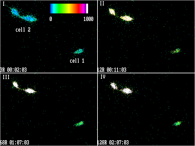
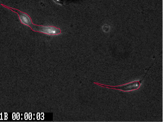
Figure 1C.
A phase-contrast image of the cells analyzed in B, photographed under dim visible light and 380 nM illumination, is shown with the outline for integration of the fluorescence emission-ratio signal.