![]() Figure 1 of
Shih, Mol Vis 4:4, 1998.
Figure 1 of
Shih, Mol Vis 4:4, 1998.
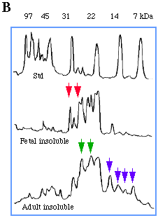
Figure 1. One-dimensional electrophoretic analysis of fetal and adult bovine lens proteins
(A) SDS-PAGE of water-soluble and water-insoluble bovine lens proteins. F= fetal, A = adult.
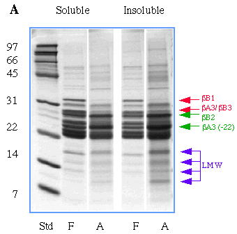
(B) Densitometric scan of lanes in (A) containing molecular weight standards, water-insoluble protein from fetal lenses, and water-insoluble protein from adult lenses. Red arrows indicate the positions of intact ßB1 and comigrating ßA3/ßB3 crystallins which decrease with age. Green arrows indicate intact ßB2 crystallin and partially degraded ßA3 crystallin missing 22 residues from its N-terminus which increase with age. Purple arrows show positions of extensively degraded crystallins found in highest concentration in the insoluble protein of adult lenses. The identity of crystallins in this figure were inferred based on their apparent molecular weights and comparison to proteins identified on two-dimensional electrophoretic gels shown in Figure 2.