![]() Figure 9 of
Nickerson, Mol Vis 4:33, 1998.
Figure 9 of
Nickerson, Mol Vis 4:33, 1998.
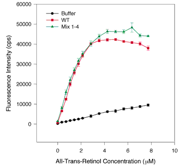
Figure 9. Comparison of WT and an equimolar mix of the four repeats by fluorescence enhancement of all-trans-retinol
One µM WT protein in 700 µl was added to one cuvette and a mix consisting of 1 µM of each repeat was added to another cuvette. The same assay conditions were employed as in Figure 1. Retinol fluorescence enhancement was measured by excitation at 339 nm and emission at 479 nm. The error bars indicate the standard error of the mean. The intact WT IRBP is indicated by the red squares, and the green triangles signify the mixture of the four repeats. The buffer alone is shown by the black circles. The mixture of four repeats displays similar, but not identical, binding behavior to WT intact protein.
