![]() Figure 2 of
Nickerson, Mol Vis 4:33, 1998.
Figure 2 of
Nickerson, Mol Vis 4:33, 1998.
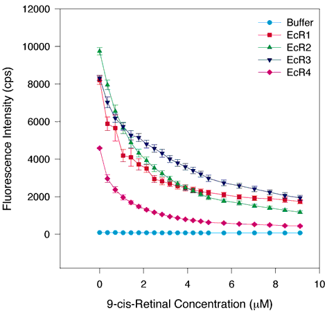
Figure 2. 9-cis-Retinal quenching of tryptophan quenching of fluorescence in WT and individual repeats of IRBP
The same assay conditions were employed as in Figure 1. 9-cis-retinal also quenches tryptophan fluorescence in all four repeats. Analysis of the binding curves suggests that 9-cis-retinal binds to each repeat with high affinity and the binding is saturable, indicating a specific binding of ligand and receptors. The binding curve parameters all differ slightly suggesting a general similarity of properties of the four individual repeats. The symbols and colors are the same as in Figure 1. The error bars indicate the standard error of the mean.
