![]() Figure 6 of
He, Mol Vis 4:32, 1998.
Figure 6 of
He, Mol Vis 4:32, 1998.
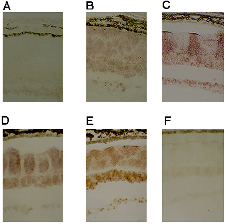
Figure 6. Determination of the spatiotemporal expression of the Nrl gene in developing rat retinas using in situ hybridization.
The expression of Nrl gene in light-adapted rat retinas at (A) PN5, (B) PN10, (C) PN15, (D) PN21, and (E) PN45 was detected using biotin-labeled oligo probes and immunogold-silver enhancement techniques. The Nrl gene expression was detectable at PN5, mostly in the inner nuclear (INL) and ganglion cell (GCL) layers. The expression of Nrl increased during development, reaching a peak at PN15. Note that the pattern of Nrl expression shifted from the proximal to distal or outer retina throughout development. Figure 6F is a PN45 retina hybridized with sense probe, which showed no signal at all. Similarly, no signal was observed on slides incubated in the absence of Nrl, colloidal gold-streptavidin or silver enhancement reagent (data not shown). The expression of Nrl in light-adapted developing and adult retinas was not different from that in dark-adapted retinas (data not shown).