Figure 5. Ophthalmic examination instruments reveal the clinical manifestations of illustrative cases. A: Anterior segment photograph of the right eye of illustrative case 1 showed mild cortical cataract. B: Anterior segment photograph of the left eye of illustrative case 1 showed corneal pannus. C: UBM photograph of the right eye of illustrative case 1 showed open angle. D: UBM photograph of the left eye of illustrative case 1 showed open angle. E: B-scan ultrasound of the right eye of illustrative case 2 indicated a large cup. F: B-scan ultrasound of the left eye of illustrative case 2 indicated normal result. G: UBM photograph of the right eye of illustrative case 2 showed closed angle. H: UBM photograph of the left eye of illustrative case 2 showed open angle.
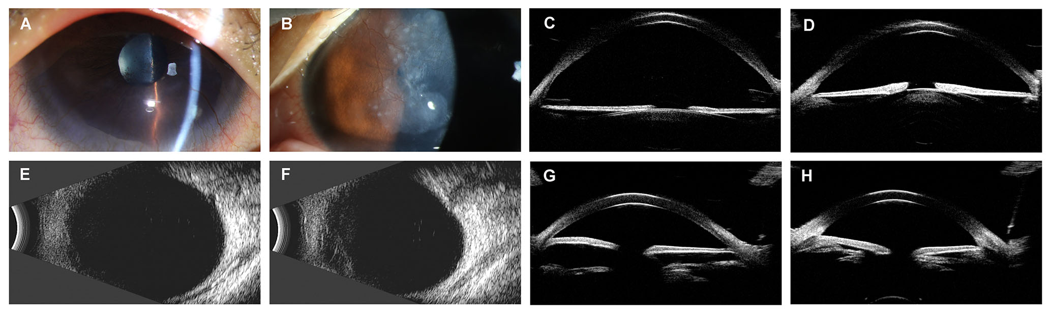
 Figure 5 of
Wang, Mol Vis 2025; 31:221-229.
Figure 5 of
Wang, Mol Vis 2025; 31:221-229.