![]() Figure 2 of
Ban, Mol Vis 3:18, 1997.
Figure 2 of
Ban, Mol Vis 3:18, 1997.
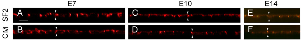
Figure 2. RPE forms monolayers with ZO-1 and actin in apical junctional complexes.
Cultured RPE formed monolayers of similar height with ZO-1 and actin colocalized in apical junctional complexes. The cultures described in Figure 1 were examined by confocal microscopy in the transverse plane. In addition to apical, circumferential bands of actin, a small population of actin filaments was dispersed through out the cytoplasm. Actin staining (A,B,C,D) was used to determine cell height and for evidence of contaminating fibroblasts. In all fields examined (from triplicate filters, and from 4-6 experiments), there was no evidence of multilayering of RPE or an underlying layer of contaminating fibroblasts. ZO-1 colocalized with the circumferential bands of actin, as exemplified by double labeled cells on E14 (E and F). There was no significant difference in cell height between cultures. Downward arrows, apical membrane; Upward arrows, basal membrane; Bar, 10 µm.