![]() Figure 1 of
Ban, Mol Vis 3:18, 1997.
Figure 1 of
Ban, Mol Vis 3:18, 1997.
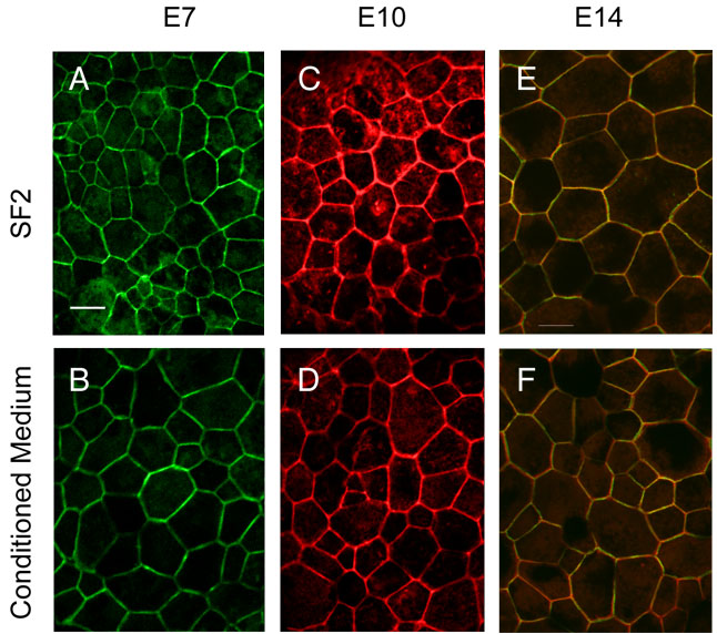
Figure 1. ZO-1 and actin colocalize at cell-cell junctions.
Cultured RPE formed hexagonal arrays, and filamentous actin overlapped with ZO-1 at cell-cell junctions. RPE was isolated from E7, E10 or E14 embryos and cultured for 5 days on laminin-coated filters. Cultures were maintained in SF2 growth medium (A,C,E) or stimulated with E14 retinal conditioned medium (B,D,F). ZO-1 and actin were localized by indirect immunofluorescence using antibody R40.76 (ZO-1), or fluorescently tagged phalloidin (actin). Confocal images were obtained in an en face plane near the apical membrane. Similar results were obtained after 10 days in culture and with polyclonal antibody 7445 to ZO-1. Examples are shown for ZO-1 label (A and B), actin label (C and D) and double label (E and F). There was no apparent effect of conditioned medium. Bar, 10 µm.