Figure 5. Simulated three-dimensional crystal structures of proteins. Predicted crystal structures of wildtype (left) and mutant (right)
proteins. Yellow: wildtype (left) and mutant (right) residues. Green: residues that bind to wildtype (left) and mutant (right)
residues.
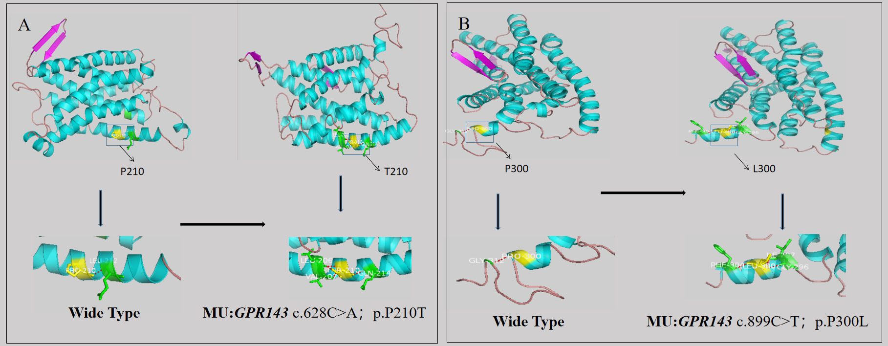
 Figure 5 of
Xu, Mol Vis 2023; 29:234-244.
Figure 5 of
Xu, Mol Vis 2023; 29:234-244.