Figure 2 of
Xu, Mol Vis 2023; 29:234-244.
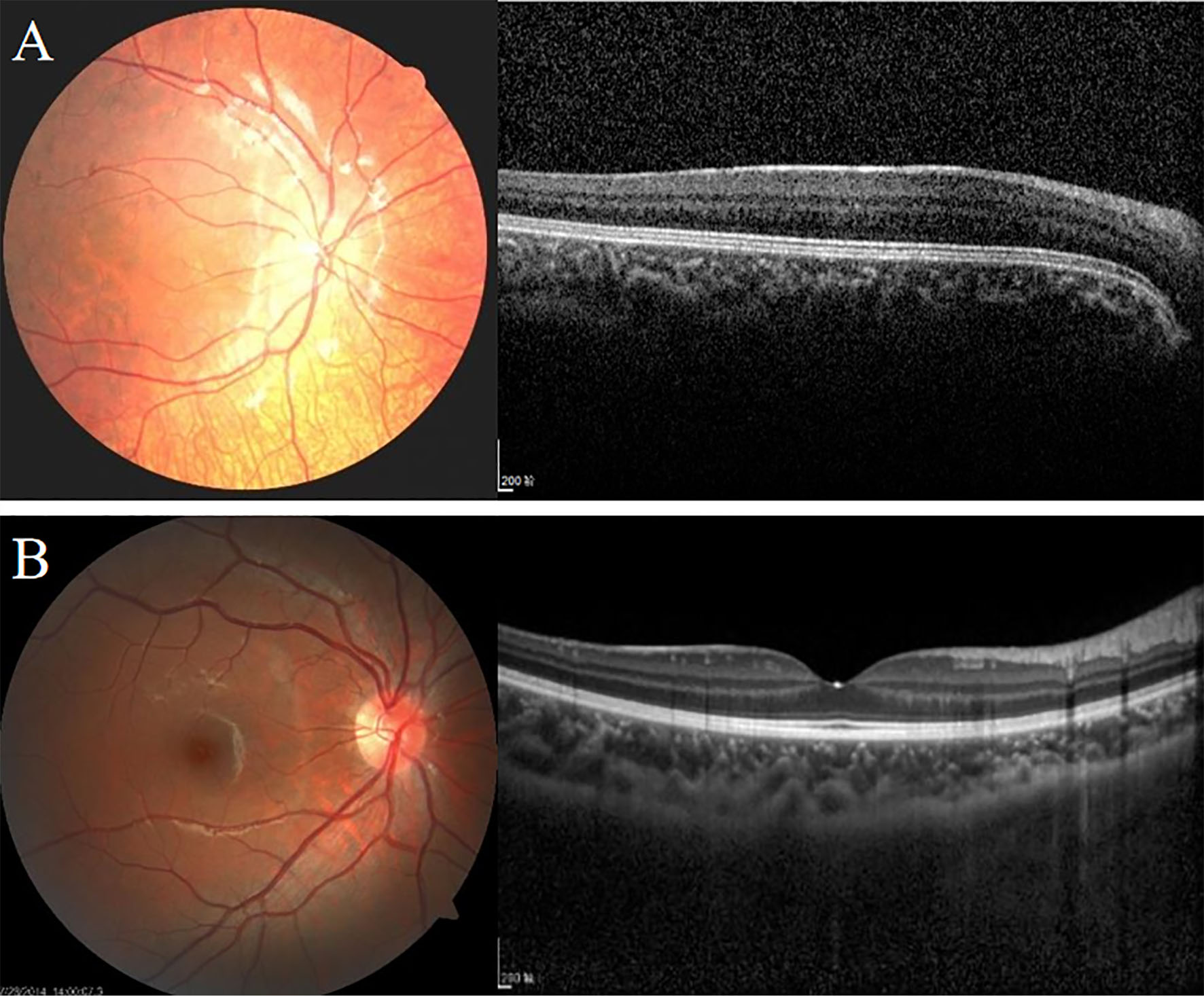
Figure 2.
Images generated from the fundus camera and SD-OCT stack. Macular OCT images showing the fundus appearance in (
A
) patients with ocular albinism type 1 (OA1) and (
B
) healthy people.
 Figure 2 of
Xu, Mol Vis 2023; 29:234-244.
Figure 2 of
Xu, Mol Vis 2023; 29:234-244.