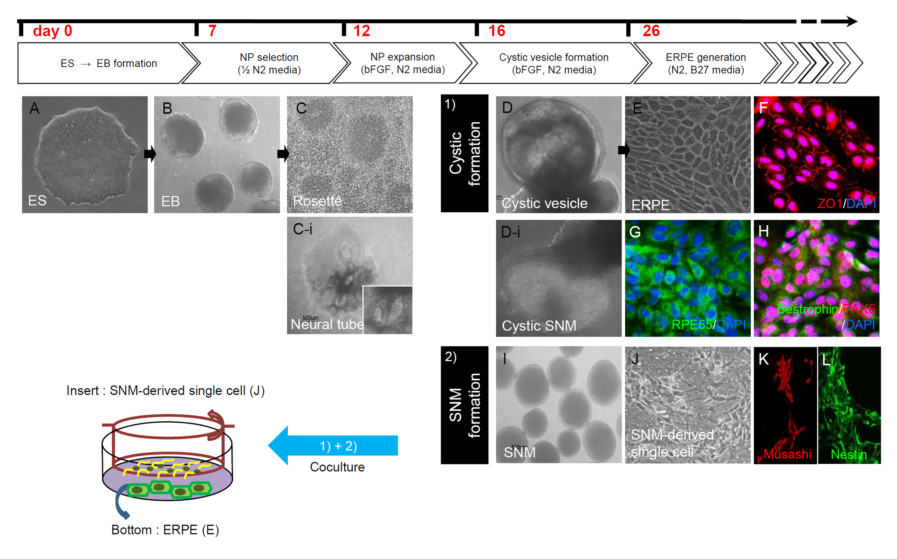
Figure 1. Figure 1. Differentiation of photoreceptor precursors from human embryonic stem (ES) cells via embryonic body (EB) and spherical neural
masses (SNMs). Human ES cells (
A) are detached to generate EBs (
B), which were then attached and cultured in neural precursor (NP) selection medium including 0.5% N2 supplement for 5 days.
Then, the medium was switched to NP expansion medium with 1% N2 supplement and basic fibroblast growth factor (bFGF). Neural
rosettes (
C) and neural tube structures (
C-
I) were mechanically isolated and then cultured in suspension to form SNMs. Some SNMs in early culture show cystic structures
(
D) that balloon outward, and these cystic SNMs (
D-
I) were separated and cystic portions were fragmented with mechanical dissection under a stereomicroscope and were attached
and cultured in RPE (retinal pigment epithelium) generation medium with 1% N2 supplement and 1% B-27 supplement, to form ES-derived
RPE cells (ERPE,
E), which are positive for zonula occludens 1 (ZO1,
F), retinal pigmented 486 epithelium-specific 65-kDa protein (RPE65,
G), and bestrophin (
H). Other non-cystic SNM (
I) were passaged and mechanically cut and plated as SNM-derived single cells (
J), positive for neural stem cell markers musashi (
K) and nestin (
L). SNM-derived single cells (
J) were cocultured with ES-derived RPE cells (ERPE,
E) to differentiate photoreceptor precursor cells.
 Figure 1 of
Shin, Mol Vis 2021; 27:288-299.
Figure 1 of
Shin, Mol Vis 2021; 27:288-299.