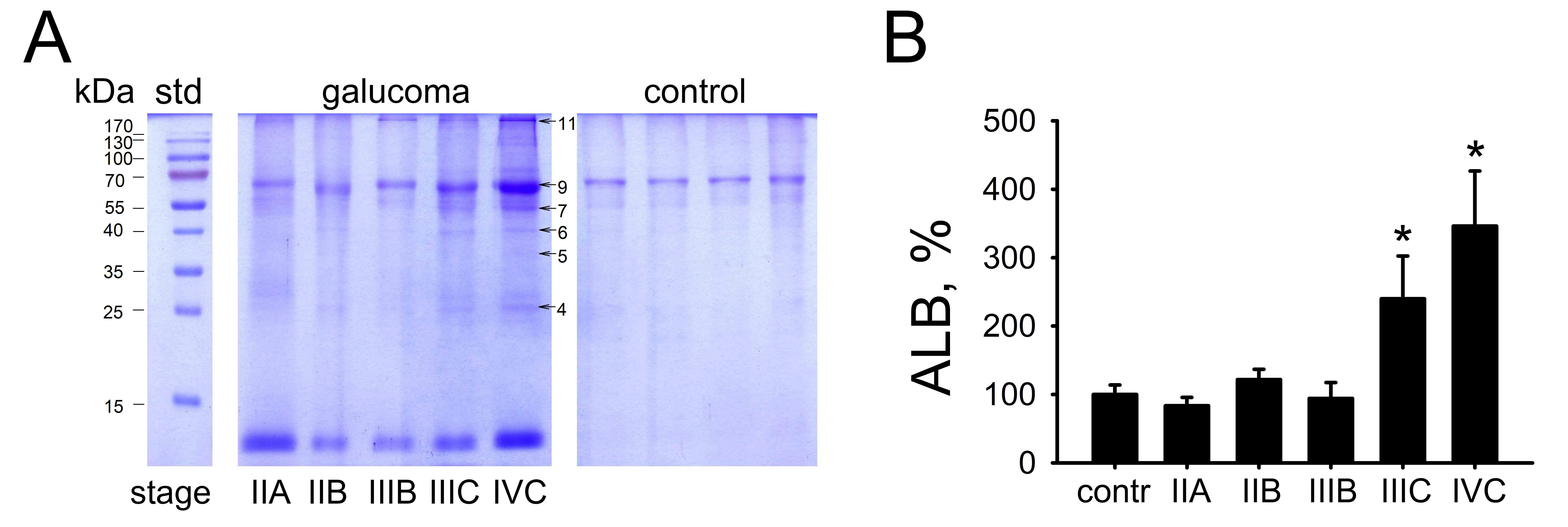
Figure 2. Determination of POAG-associated alterations in the content of the major proteins of the human sclera.
A: Representative sodium dodecyl sulfate–polyacrylamide gel electrophoresis (SDS–PAGE) images of protein extracts obtained
from non-glaucomatous sclera (control) and sclera of patients with different stages of primary open-angle glaucoma (POAG;
glaucoma). The amount of the total protein in each track is adjusted using glyceraldehyde-3-phosphate dehydrogenase (GAPDH)
as a loading control in parallel western blotting experiments (not shown). Protein standards in kDa (track std) are denoted
in the left column. Arrows indicate six proteins (the protein numbers are the same as in
Figure 1) exhibiting altered content in the sclera of the patients with POAG.
B: The weight fractions of serum albumin (ALB) estimated from the SDS–PAGE data obtained for control individuals (100%) and
the patients with different stages of POAG. *p<0.05 compared to the values obtained for the control group.
 Figure 2 of
Iomdina, Mol Vis 2020; 26:623-640.
Figure 2 of
Iomdina, Mol Vis 2020; 26:623-640.