 Appendix 5 of
Zhang, Mol Vis 2019; 25:462-476.
Appendix 5 of
Zhang, Mol Vis 2019; 25:462-476. Appendix 5 of
Zhang, Mol Vis 2019; 25:462-476.
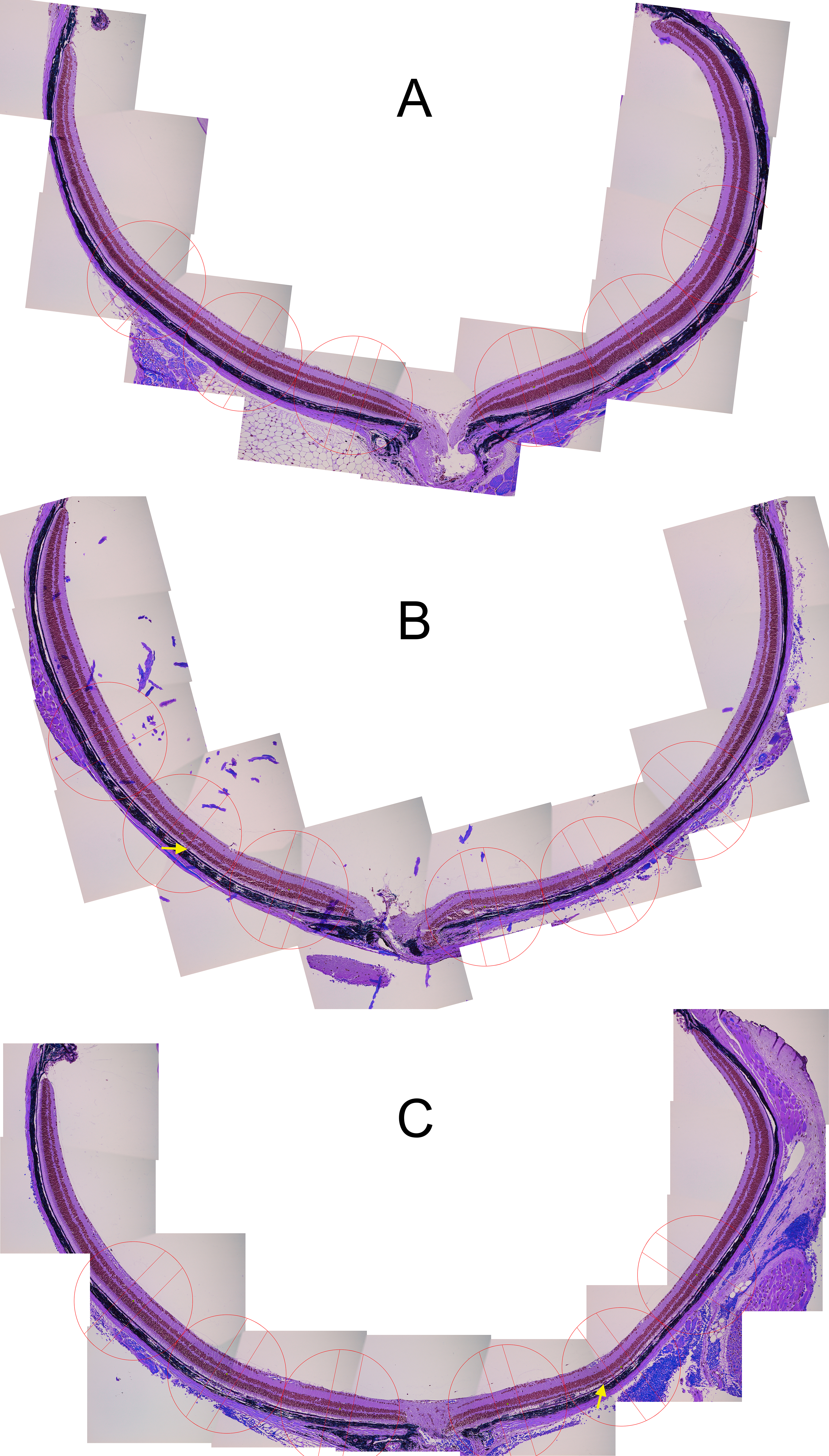
Appendix 5.
Figure showing full-sized images that suggest that exercise partially preserves nuclei of the outer nuclear layer in the I307N Rho retinal degeneration. The I307N Rho degeneration was induced in active and inactive mice. A week later the mice were euthanized and ocular sections stained with hematoxylin and eosin (H&E). Representative H&E images of complete retina sections from each treatment group are shown with six spacing and counting overlay templates per section as used for quantification of nuclei of the outer nuclear layer starting at 250 µm from the optic nerve head and extending every 500 µm outward along both the dorsal/superior (right half of each section) and ventral periphery/inferior (left half of each section). The same set of high-resolution images of complete retina sections were used for quantification of nucleated cell infiltrates observed the interphotoreceptor space (quantification presented in Figure 8). Examples of nucleated infitrates are pointed to by arrows. A: Section from an inactive, uninduced mouse shows unperturbed ONL across the span of the retina. B: Section from an inactive, induced mouse shows extreme thinning in the central, superior (right side of section) ONL. C: Section from an active, induced mouse shows diminished thinning ONL relative to inactive mouse. To access the data, click or select the words “Appendix 5.”