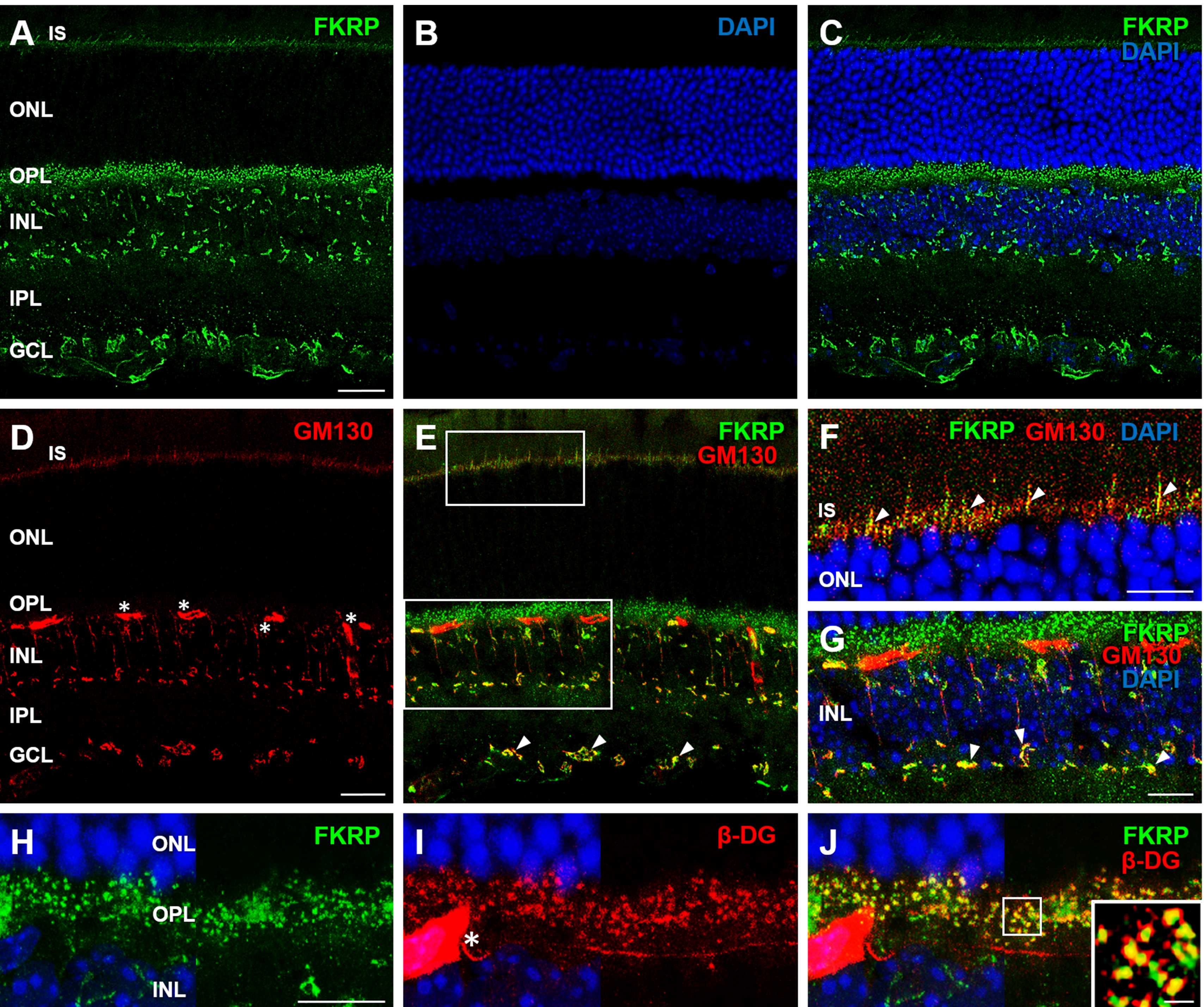
Figure 4. Immunolocalization of FKRP in the mouse retina. Double immunostainings were performed on mouse retinal sections for FKRP (
A,
C,
E,
H,
J; green) and the Golgi complex marker GM130 (
D,
E; red) or β-dystroglycan (β-DG;
I,
J; red). Enlarged views in
F and
G correspond to boxed areas in
E, and the boxed area in
J is shown enlarged in the lower right inset. Nuclei stained with DAPI are shown in blue (
B,
C,
F-J). FKRP showed immunoreactivity in the photoreceptor IS and in the OPL, INL and GCL (
A,
C,
E). The double immunolabeling for FKRP and GM130 revealed colocalization in the photoreceptor IS, INL and GCL (
E-
G, arrowheads), whereas the double immunostaining for FKRP and β-DG showed colocalization in the OPL (
J; yellow). Asterisks in
D and
I indicate non-specific immunostaining of retinal vessels by secondary anti-mouse IgG. Abbreviations are given in the legend
of
Figure 2. Each bar equals 20 μm (
A-
E), 10 μm (
F-
I) or 1 μm (
J, inset).
 Figure 4 of
Haro, Mol Vis 2018; 24:43-58.
Figure 4 of
Haro, Mol Vis 2018; 24:43-58.