Figure 2. Immunolocalization of fukutin in the mouse retina. Double immunostainings were performed on mouse retinal sections for fukutin
(A, C, E, I; green), the specific Golgi complex marker GM130 (D, E; red), and the specific ER marker calreticulin (CALR; H, I; red). Enlarged views in F and G correspond to boxed areas in E, and enlarged views in J and K to boxed areas in I. Nuclei stained with DAPI are shown in blue (B, C, F, G, J and K). Fukutin immunoreactivity was found in the cytoplasm and nucleus of cells in the INL and the GCL of the mouse retina, but
without colocalizing with DAPI (C, arrowheads). However, colocalization of fukutin with calreticulin was observed in the IS of photoreceptors and the OPL (I-K), but without noticeable colocalization with GM130 (E-G). Asterisks in D and H indicate non-specific immunostaining of retinal vessels with secondary antibodies to mouse IgG. IS=inner segments; ONL=outer
nuclear layer; OPL=outer plexiform layer; INL=inner nuclear layer; IPL=inner plexiform layer; GCL=ganglion cell layer. Each
bar equals 20 μm, except in F, G, J and K: 10 μm.
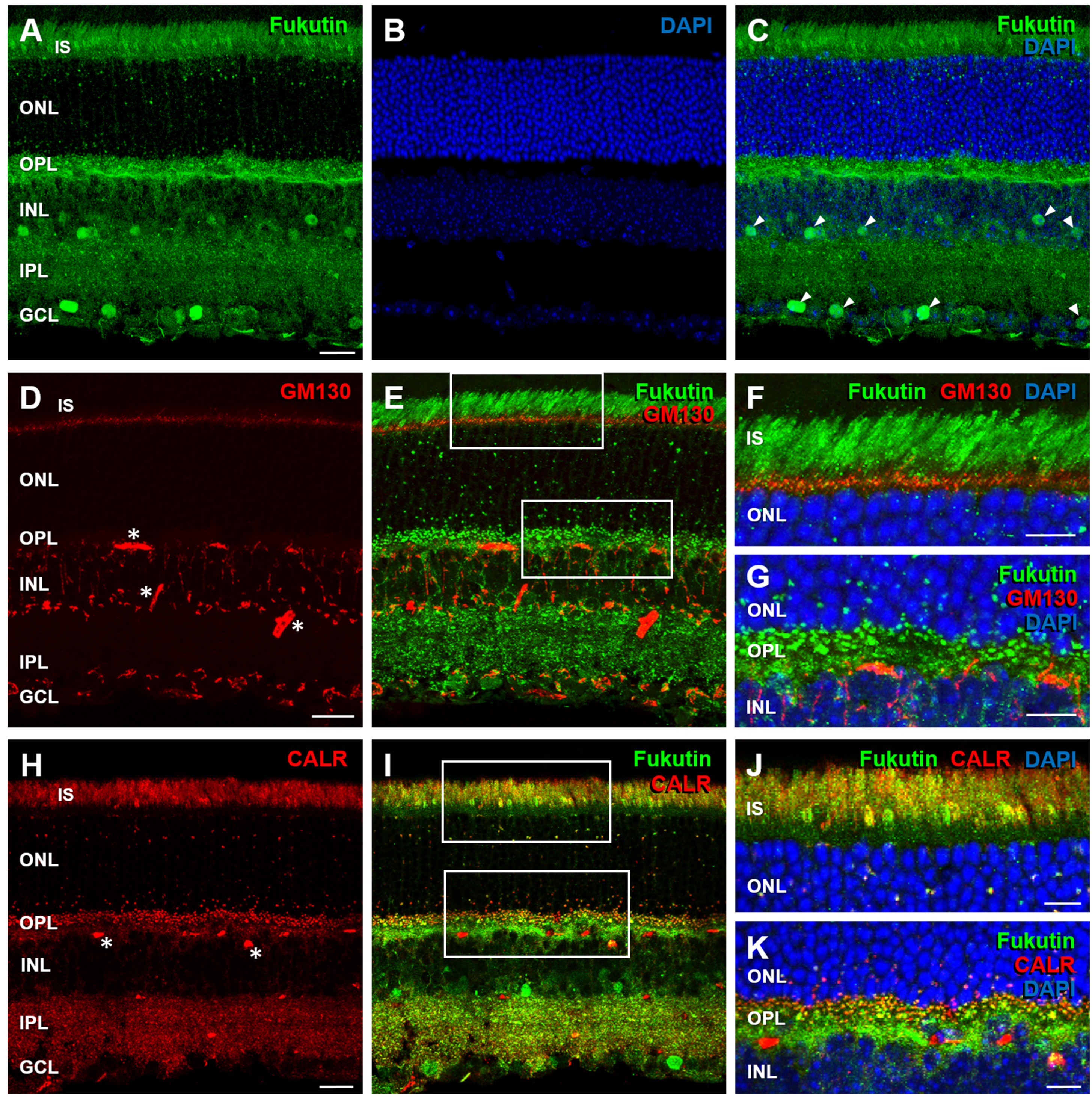
 Figure 2 of
Haro, Mol Vis 2018; 24:43-58.
Figure 2 of
Haro, Mol Vis 2018; 24:43-58.