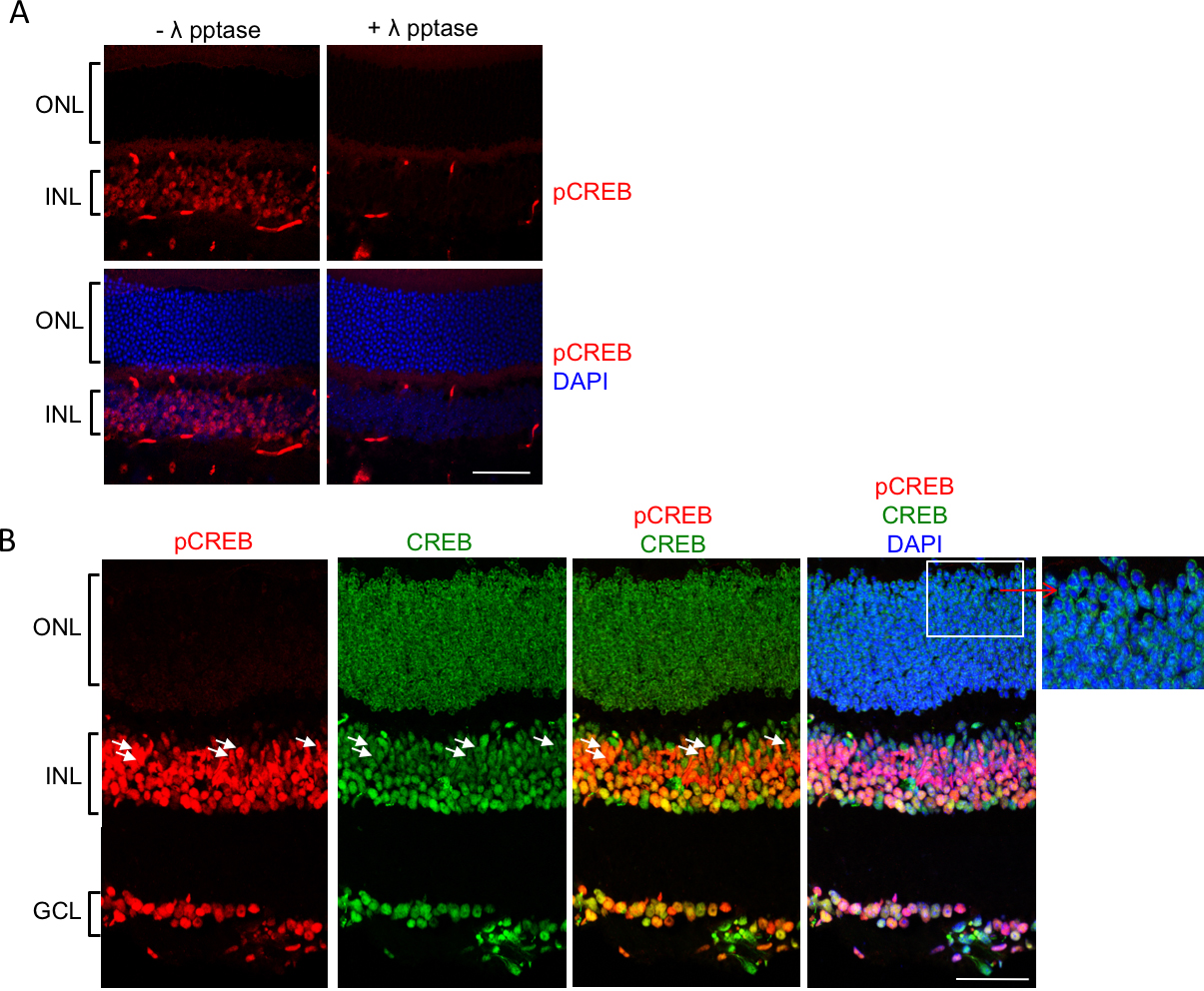
Figure 2. Expression and localization of CREB and pCREB in the wild-type retina at P28.
A: To verify that phosphorylated cyclic AMP response element binding protein (pCREB) is selectively recognized by the anti-pCREB
antibody, wild-type retina sections from mice at P28 were treated without (-) and with (+) λ phosphatase (λ pptase) as described
in the Methods, followed by staining with anti-pCREB conjugated to Alexa Fluor 488A (anti-pCREB-Alexa Fluor 488 conjugate;
1:25). pCREB staining was then pseudocolored red using Adobe Photoshop to be consistent with
Figure 2B. No staining was detected in the sections treated with λ phosphatase, indicating that the antibody is specific for the phosphorylated
form of CREB.
B: CREB (green) expression is evident in all three nuclear layers. In contrast, pCREB (stained with the unconjugated anti-pCREB
antibody followed by secondary antibody; red) is visualized in a subset of cells, including the Müller glia (MG; based on
the elongated shape of their cell bodies; white arrows) in the INL as well as other cells in the INL and the GCL. Notably,
pCREB is absent from the ONL, which contains the nuclei of the photoreceptors.
Inset: Magnified view of the ONL; anti-CREB, 1:50; anti-pCREB, 1:200; secondary antibodies Alexa Fluor 488 goat anti-mouse immunoglobulin
G (IgG) and Alexa Fluor 555 goat anti-rabbit IgG at 1:1,000. The z-stacks from a single mouse retina were processed as maximum
projections using Adobe Photoshop. GCL, ganglion cell layer; INL, inner nuclear layer; ONL, outer nuclear layer. Scale bar
= 50 μm.
 Figure 2 of
Dong, Mol Vis 2017; 23:90-102.
Figure 2 of
Dong, Mol Vis 2017; 23:90-102.