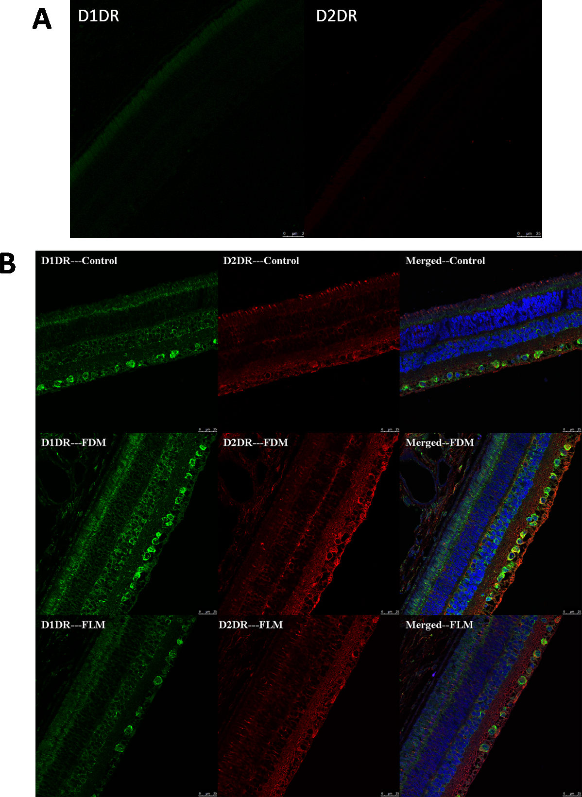
Figure 4. Photographs of retinas stained by immunofluorescence staining of D1 dopamine (DA) receptors (D1DR) and D2 DA receptors (D2DR).
Green staining is D1DR, red staining is D2DR, blue staining is the cell nucleus (scale bar: 25 μm). D1DR was mainly found
on the retinal ganglion cells (RGCs) and inner nuclear layers (INLs), while D2DR was mainly found on the RGCs and inner plexiform
layers (IPL;
Figure 4A shows the primary control images for each antibody).
 Figure 4 of
Luo, Mol Vis 2017; 23:666-679.
Figure 4 of
Luo, Mol Vis 2017; 23:666-679.