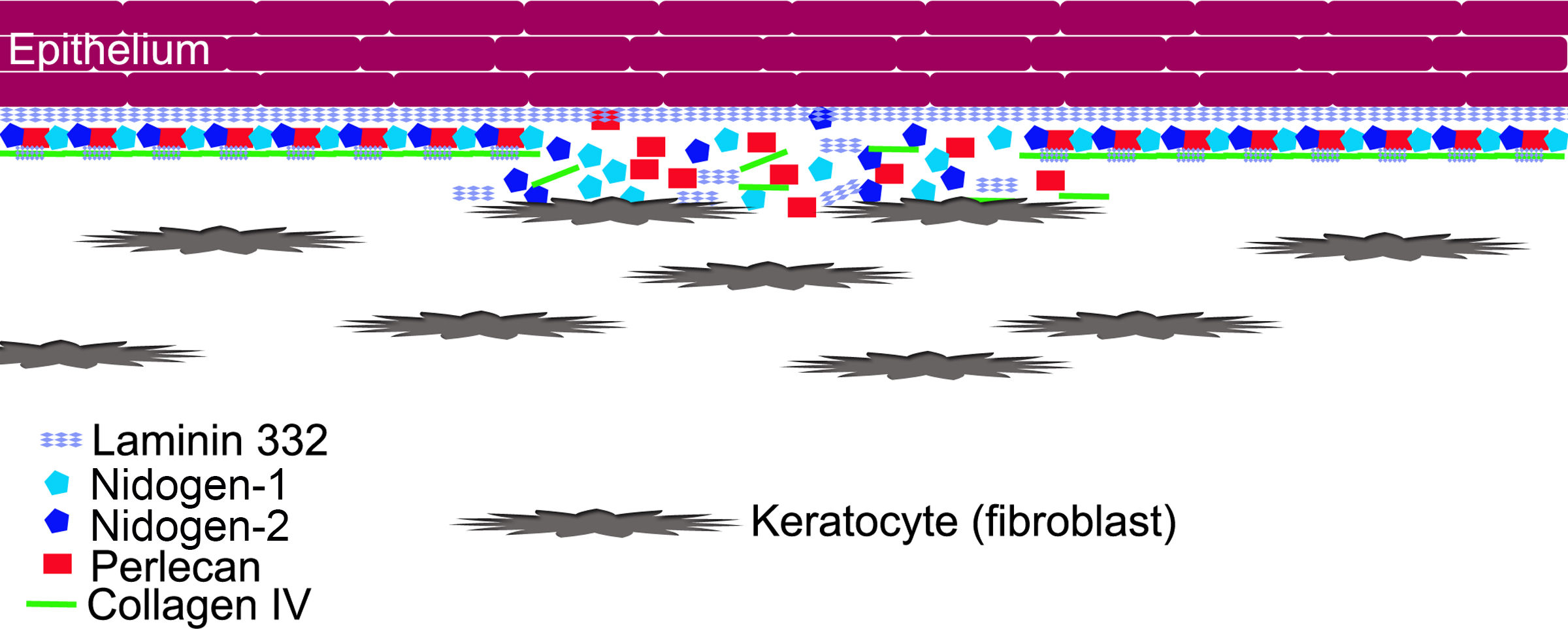
Figure 7. Schematic diagram illustrating hypothesized keratocyte contributions to EBM regeneration. We hypothesize that after corneal
epithelial and epithelial basement membrane (EBM) injury, the epithelial defect heals, and the epithelium lays down self-polymerizing
laminin 332 to form a nascent EBM. Once this layer is laid down, it forms a barrier that limits to some extent posterior penetration
of the other EBM components needed to produce the structurally and functionally mature more posterior basement membrane that
includes the lamina lucida and the lamina densa. These components must be provided by contiguous keratocytes that possibly
need to be in direct contact with the nascent EBM based on the morphology and ultrastructure noted with TEM in
Figure 1A–C. These components provided by keratocytes could include laminin chains (such as laminin α-3), nidogens, perlecan, collagens
(such as collagen type IV), and other components. In the rabbit PRK model, the laminin α-3 and nidogen-2 mRNAs were decreased
in the anterior stroma of the corneas that had −9.0D photorefractive keratectomy (PRK) compared to the corneas that had −4.5D
PRK during the days just before the appearance of the normal regenerated EBM in the corneas that do not develop fibrosis.
 Figure 7 of
Santhanam, Mol Vis 2017; 23:39-51.
Figure 7 of
Santhanam, Mol Vis 2017; 23:39-51.