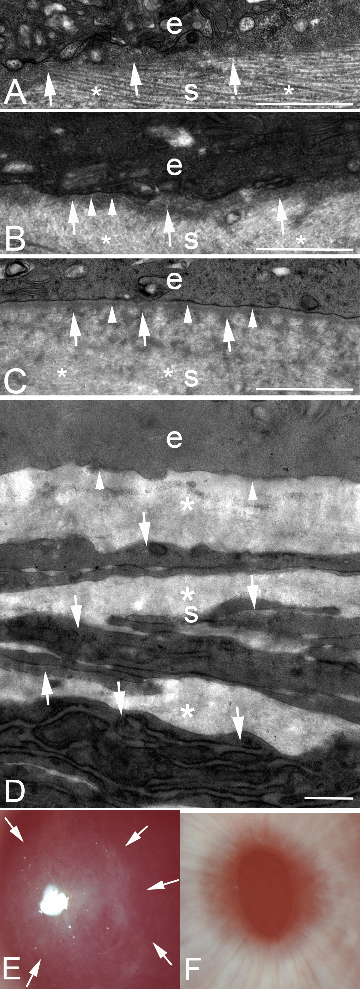
Figure 1. Transmission electron microscopy of the junction between the epithelium and the stroma at different time points after low-correction
−4.5D photorefractive keratectomy (PRK) or high-correction −9.0D PRK in rabbits. e, epithelium; s, stroma.
A: In a cornea at 7 days after −4.5D PRK, no discernable lamina lucida or lamina densa is present. Note, by chance, the specimen
was cut such that the stacked collagen lamellas (*) are seen in the stroma. Dense deposits of extracellular matrix (arrows)
can be seen in the stroma just posterior to the epithelium. Magnification = 23,000X.
B: In a cornea at 8 days after −4.5D PRK, most of the excimer laser-ablated zone had no lamina lucida or lamina densa. However,
in one area (arrowheads) nascent lamina lucida and lamina densa can be noted. Again, dense extracellular matrix seen in the
stroma just posterior to the epithelium. In the stroma, collagen lamellas (*) that have been cut transversely can be noted.
Magnification = 23,000X.
C: In a cornea at 9 days after −4.5D PRK, there is a complete lamina lucida and lamina densa (arrowheads) across 100% of the
excimer laser-ablated zone. Again, the dense extracellular matrix is noted just posterior to the intact epithelial basement
membrane (EBM). In the stroma, collagen lamellas (*) that have been cut transversely can be noted.
D: In a cornea at 1 month after −9.0D PRK that developed severe stromal fibrosis (haze), no normal EBM is detected beneath
the epithelium (arrowheads). The anterior stroma is filled with stacked myofibroblasts (arrows) with large amounts of intracellular
rough endoplasmic reticulum and surrounding disorganized extracellular matrix (*) that are the alpha-smooth muscle action+
(α-SMA) cells noted in
Figure 2A. Magnification = 23,000X.
E: Slit-lamp photograph of a rabbit cornea at 1 month after −9.0D PRK. Note the dense haze (arrows) in the central excimer
laser-treated cornea with the pupil dilated. Magnification = 40X.
F: Slit-lamp photograph of a rabbit cornea at 1 month after −4.5D PRK. Note the cornea is clear without fibrosis, and thus,
the iris details are clear. Magnification = 40X. Scale bars (
A–
D) = 2 µm.
 Figure 1 of
Santhanam, Mol Vis 2017; 23:39-51.
Figure 1 of
Santhanam, Mol Vis 2017; 23:39-51.