Figure 2. LCM strategy. A: Representative cornea stained for alpha-smooth muscle actin (α-SMA) and 4',6-diamidino-2-phenylindole (DAPI) at 1 month
after −9.0D PRK. The indicated α-SMA+ myofibroblast layer was 36 µm thick according to the ImagePro software. This was the
minimum thickness measured in nine pilot corneas at 1 month after −9.0D photorefractive keratectomy (PRK). The nuclei of all
cells were stained with DAPI. B: Representative cornea stained for α-SMA at 1 month after −4.5D PRK. Note that no α-SMA+ myofibroblasts were detected in
the stroma. The epithelium was 34 µm thick. C: Corneal section stained with Arcturus Histogene before laser capture microdissection (LCM). E, epithelium; S, stroma. D: The same corneal section after removal of two 36 µm thick specimens from the stroma immediately posterior to the epithelial
basement membrane (EBM). The areas of the two cut specimens that dropped from the laser-cut polyethylene terephthalate (PET)
membrane slide directly into a microtube with 70 µl of RNAlater RNA stabilization reagent (Ambion) is shown. Magnification = 400X.
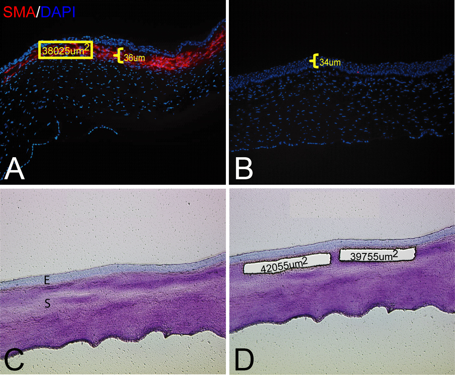
 Figure 2 of
Santhanam, Mol Vis 2017; 23:39-51.
Figure 2 of
Santhanam, Mol Vis 2017; 23:39-51.