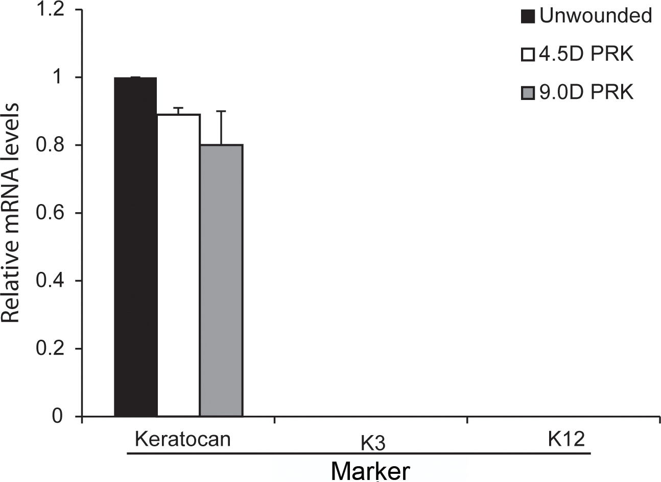
Figure 3. Verification with LCM-RT–PCR of the lack of epithelial contamination in −4.5D PRK, −9.0D PRK, or unwounded control corneas.
cDNA of laser capture microdissection (LCM) fragments cut from the anterior stroma of each cornea (as shown in
Figure 2) were analyzed using quantitative real-time PCR for keratocan (a keratocyte marker) and keratin 3 (K3) and keratin 12 (K12;
epithelial cell markers). Keratocan (keratocytes), but not K3 or K12, mRNA was detected in all three types of anterior stromal
samples. Any cDNA sample that showed K3 or K12 contamination was discarded, and cDNA was prepared from newly cut LCM fragments.
 Figure 3 of
Santhanam, Mol Vis 2017; 23:39-51.
Figure 3 of
Santhanam, Mol Vis 2017; 23:39-51.