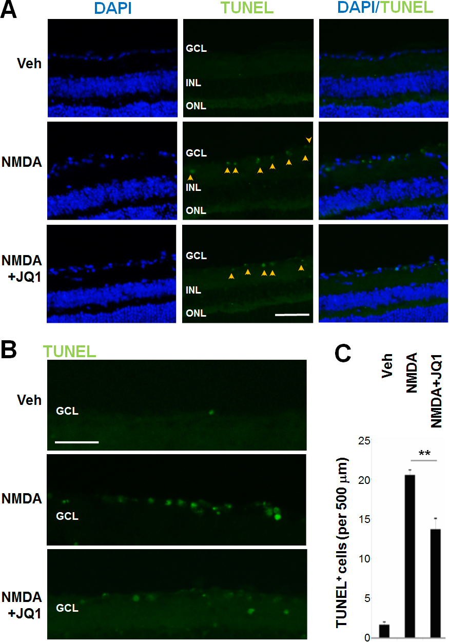
Figure 4. JQ1 attenuates NMDA-induced increase of TUNEL-positive cells. Intravitreal injections were performed as described in
Figure 1. Mice were euthanized at 24 h post injection.
A: TUNEL staining was performed on retinal cryosections. Arrowheads highlight TUNEL-positive nuclei in the GCL. Scale Bar:
50 μm.
B: Enlarged images are shown for better visibility of TUNEL-positive nuclei. Scale Bar: 50 μm.
C: Quantification: mean ± SEM of TUNEL-positive nuclei in the GCL (per 500 μm retinal length); n>6 animals; **p<0.01 compared
to NMDA alone.
 Figure 4 of
Li, Mol Vis 2017; 23:149-159.
Figure 4 of
Li, Mol Vis 2017; 23:149-159.