Figure 1. JQ1 ameliorates NMDA-induced cell loss counted in the RGC layer of retinal sections. Mice received NMDA mixed with (or without)
JQ1 in one eye and vehicle control (equivalent amount of DMSO, no NMDA) in the contralateral eye; they were euthanized at
1, 3, 7, and 14 days after injection, as described in the Methods section. Retinal cryosections were prepared for counting
DAPI-stained nuclei. A: Representative fluorescent images. Scale bar: 50 μm. GCL, retinal ganglion cell layer; INL, inner nuclear layer; ONL, outer
nuclear layer. Arrows highlight DAPI-stained nuclei in the GCL. B: Quantification of nuclei in the GCL (per 500 μm retinal length): mean ± SEM, n>6 mice; **p<0.01 compared to NMDA alone (no
JQ1).
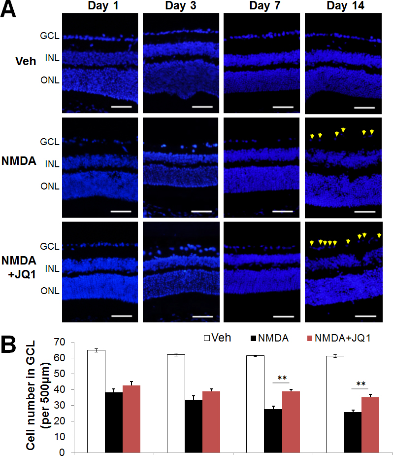
 Figure 1 of
Li, Mol Vis 2017; 23:149-159.
Figure 1 of
Li, Mol Vis 2017; 23:149-159.