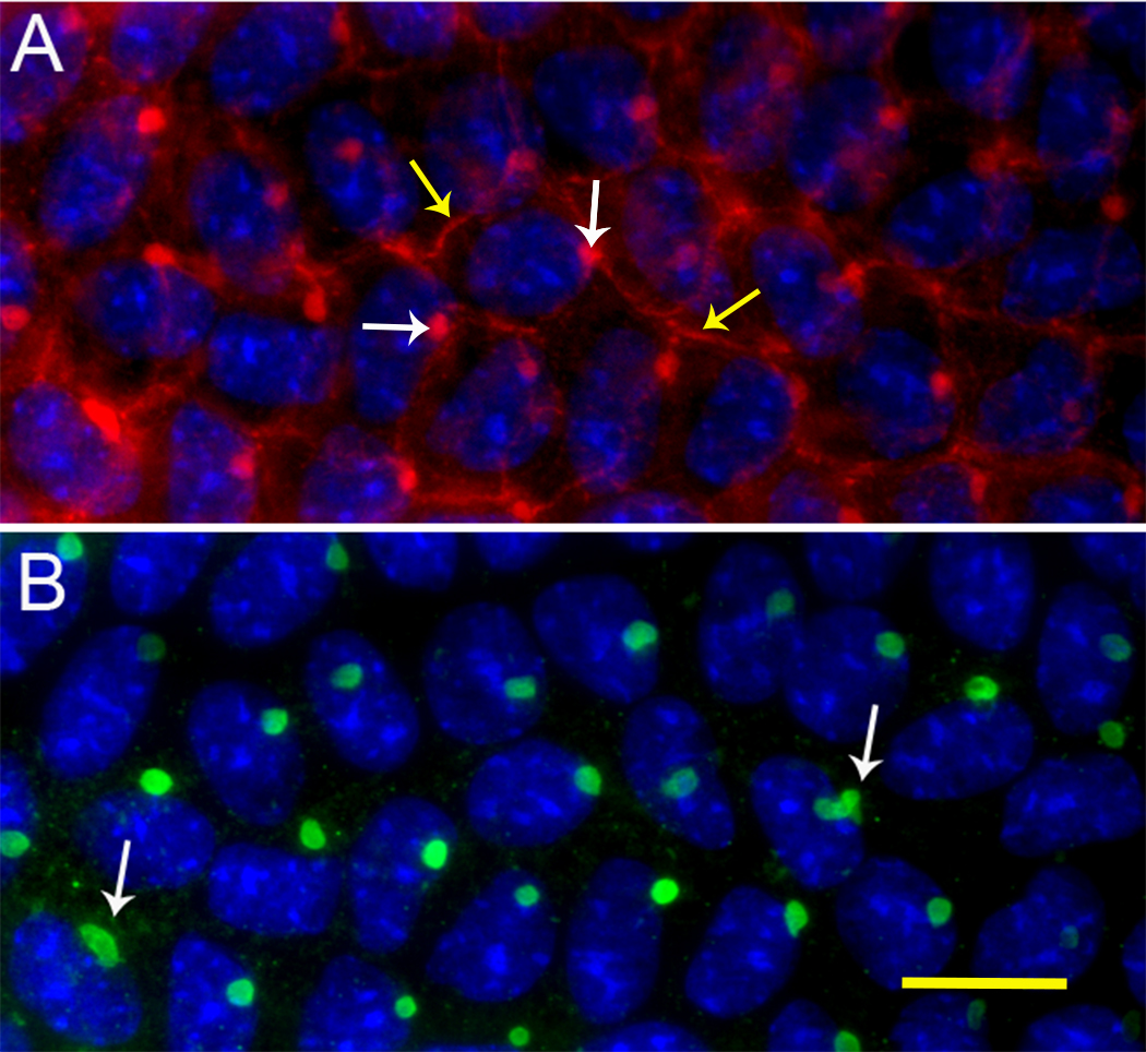
Figure 9. Localization of actin and CP49 in the vimentin knockout mouse.
A: Phalloidin-labeled filamentous actin (red channel) in an explant of lens epithelium, showing the cortical actin network
(yellow arrows), and the vermiform structure (white arrows).
B: CP49 labeling of vermiform structure in an explant of the vimentin knockout mouse. Compared to
Figure 2, it is evident that the absence of vimentin dramatically alters the shape of the vermiform structure. Labeling the vimentin
knockout reveals that the structure changes from vermiform to predominantly round. Some exceptions are usually seen, as indicated
by the white arrow in
B (scale bar = 10 μm).
 Figure 9 of
FitzGerald, Mol Vis 2016; 22:970-989.
Figure 9 of
FitzGerald, Mol Vis 2016; 22:970-989.