Figure 2. En face view of the lens epithelium. A: A lens capsule/epithelial explant labeled for CP49 (green channel) and 4',6-diamidino-2-phenylindole (DAPI; blue channel),
showing the relative uniformity of the size and distribution of this structure in the anterior epithelium (scale bar = 30
μm). B–F: Although the beaded filament (BF)-positive structure is predominantly tubular, its overall shape can vary (CP49 = green
channel). E: This structure can be present at two copies per cell (B–E scale bar = 10 μm). F: High magnification view of a fully elongated example (scale bar = 10 μm). G–H: Stimulated emission depletion (STED) super-resolution imaging of a lens epithelial explant labeled for CP49 (G, green), filensin (H, red), and merged (I). J: Deconvoluted image of colocalized CP49 and filensin showing that there may be some degree of substructure, or non-coincident
labeling of CP49 and filensin (G–H scale bar = 2 μm).
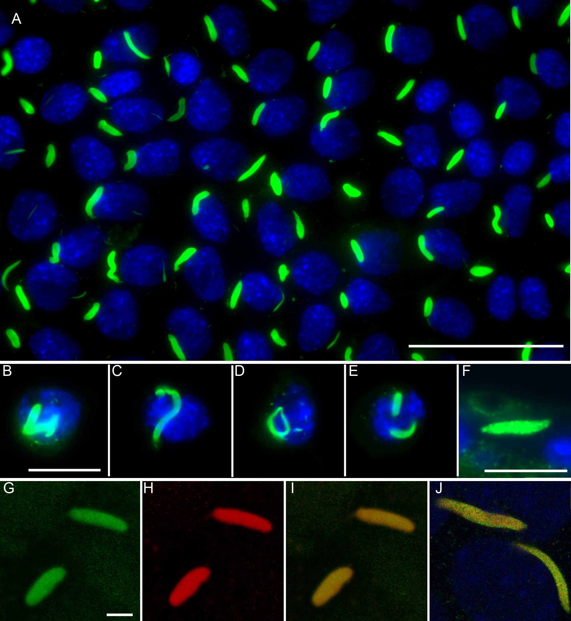
 Figure 2 of
FitzGerald, Mol Vis 2016; 22:970-989.
Figure 2 of
FitzGerald, Mol Vis 2016; 22:970-989.