Figure 5. Identification of fluorescence proteins in HEK293T cells. Protein expression of the Wt or Mut vectors was examined 48 h after
transfection. Images were captured using a 40X objective lens. A–C: Images of HEK293T cells transfected with the Wt construct encoding an enhanced green fluorescent protein that localizes
to the nucleus (A), the fusion protein of 10 aa MerTK Wt-DsRed2 is present in the cytoplasm (B). Merged images (A and B) confirm the fusion protein of the Wt construct is expressed only in the cytoplasm (C). D–F: Images of HEK293T cells transfected with the Mut construct encoding an enhanced green fluorescent protein that localizes
to the nucleus (D). E: No fluorescence fusion protein from the Mut construct was detectable in the cytoplasm. The merged images (D and E) indicate no fluorescence fusion protein expressed, except the enhanced green fluorescent protein that was observed in the
nucleus (F).
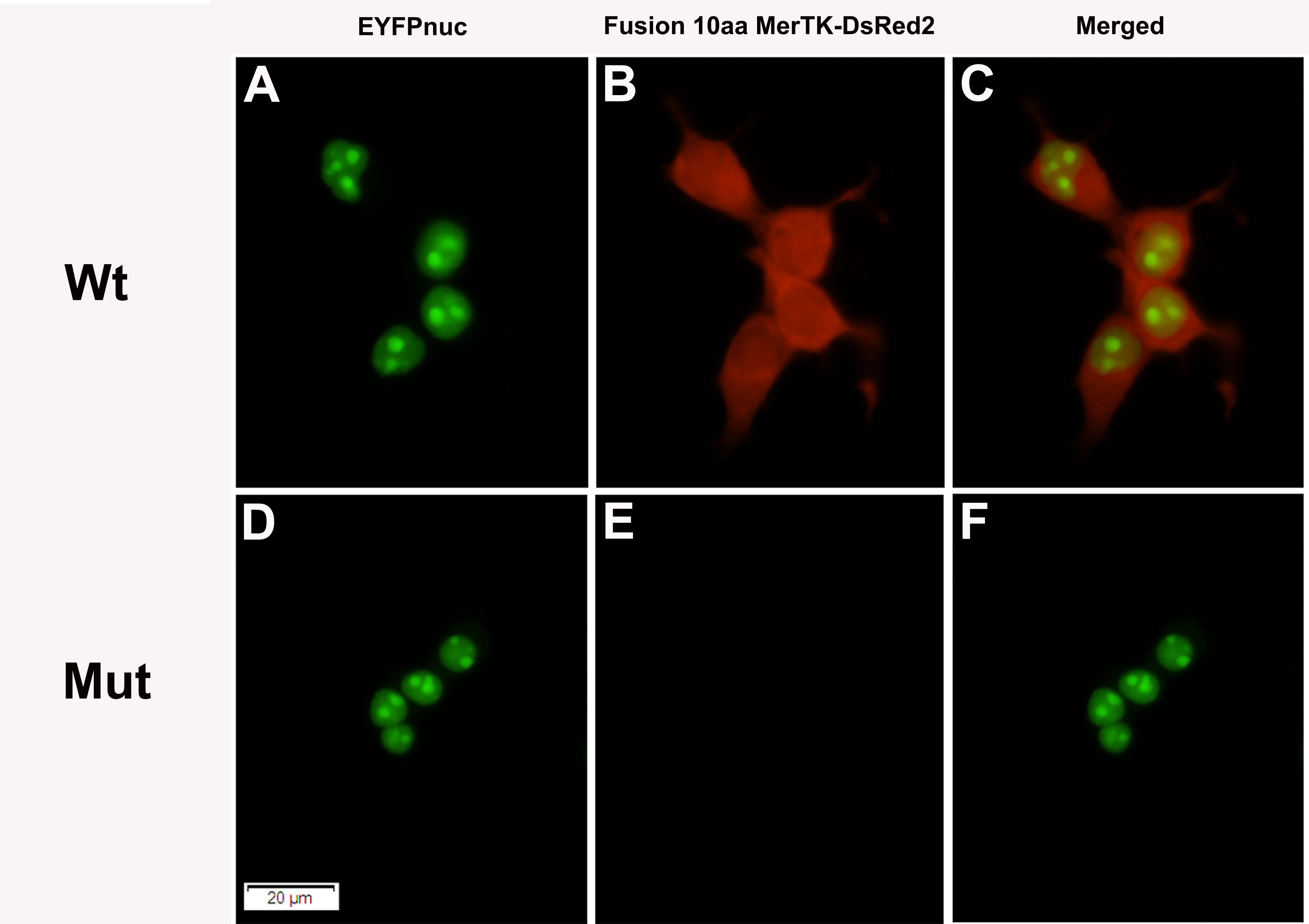
 Figure 5 of
Jinda, Mol Vis 2016; 22:342-351.
Figure 5 of
Jinda, Mol Vis 2016; 22:342-351.