Figure 1. Fundus photographs, FAF, and OCT results of the patient. Color montage fundus photographs of the right eye (A) and the left eye (B) of the patient at age 35 years. Fundus examination demonstrated changes typical of retinitis pigmentosa (RP) in both eyes,
including pale optic discs with attenuated retinal vessels, generalized pigmentary granularity, moderate bone-like spicules
in four quadrants, and macular atrophy with pigment accumulation. Fundus autofluorescence (FAF) imaging of the right eye (C) and the left eye (D) showed bilateral decreased autofluorescence corresponding to the areas of pigment accumulation in the center of the macula.
These areas were encircled by spotty autofluorescence along the temporal vascular arcades. The optical coherence tomography
(OCT) scans of the right eye (E) and the left eye (F) revealed decreased macular thickness.
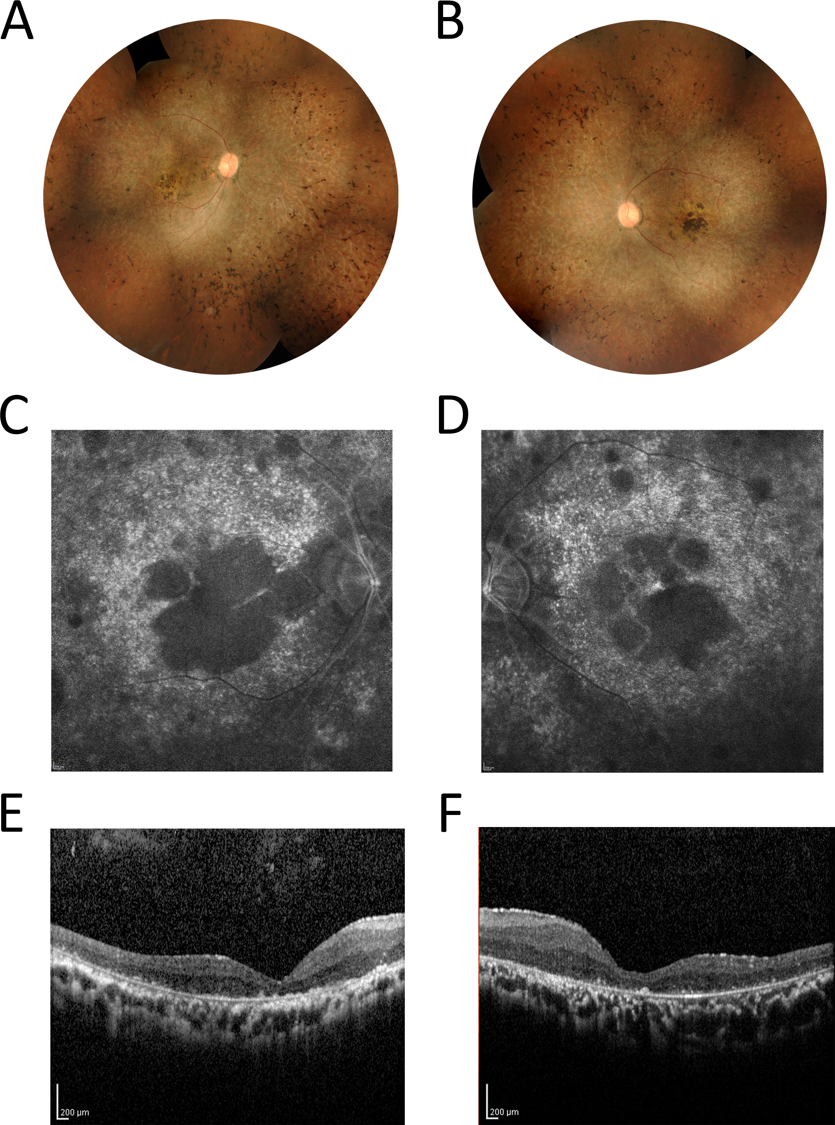
 Figure 1 of
Jinda, Mol Vis 2016; 22:342-351.
Figure 1 of
Jinda, Mol Vis 2016; 22:342-351.