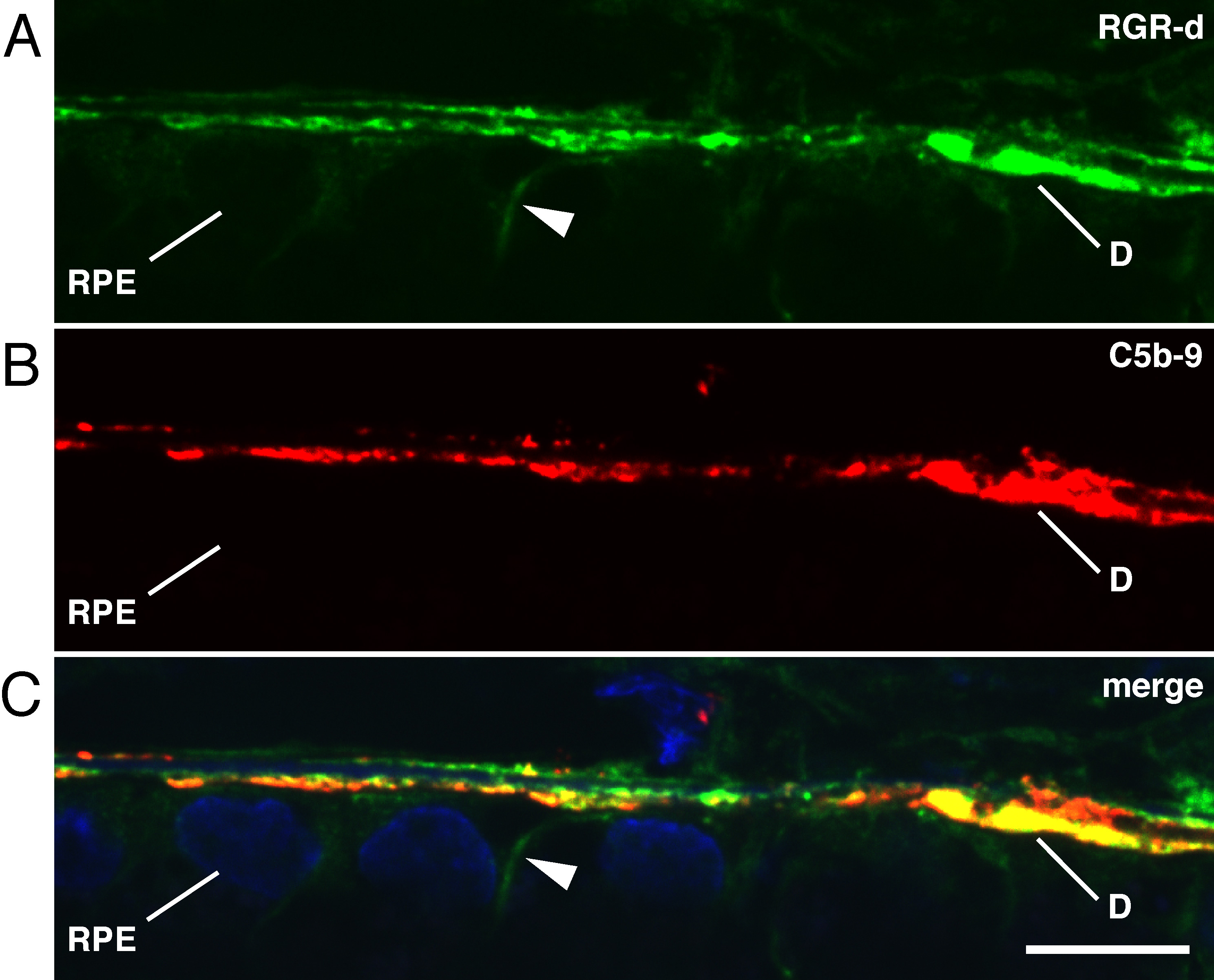
Figure 7. Localization of extracellular RGR-d and C5b-9 complement complex in basal deposits determined by double-labeling immunofluorescence
in an RPE-choroid section from a 64-year-old female (same donor as in
Figure 2).
A: Basal deposits are immunoreactive with RGR-d-specific DE21 antibody and anti-rabbit IgG conjugated to FITC.
B: Basal deposits are labeled with C5b-9 monoclonal antibody and anti-mouse IgG antibody conjugated to Cy3.
C: The merged image with DAPI counterstain shows significant co-localization of RGR-d and C5b-9 in basal deposits (
D). The arrowhead shows faint labeling of RGR-d in the basolateral plasma membrane. Scale bar, 10 μm.
 Figure 7 of
Kochounian, Mol Vis 2016; 22:213-223.
Figure 7 of
Kochounian, Mol Vis 2016; 22:213-223.