Figure 2. RGR-d and CD46 in the basolateral plasma membrane of human RPE. The RPE-choroid tissue section from a 64-year-old female donor
was incubated with RGR-d-specific DE21 antibody and a monoclonal antibody directed against CD46. Double-labeling immunofluorescence
with the FITC and Cy3 fluorochromes for RGR-d and CD46, respectively, was visualized by confocal microscopy. A: Immunofluorescent labeling of RGR-d in the RPE basolateral plasma membrane (arrowheads). Even stronger labeling was seen
in Bruch’s membrane (BrM; arrows). B: Immunofluorescent labeling of CD46 in the RPE basolateral plasma membrane (arrowheads). C: Localization of both RGR-d and CD46 in the basolateral plasma membrane. The merged image with DAPI counterstain showed no
immunostaining of CD46 in RGR-d positive areas of Bruch’s membrane. Scale bar, 10 μm.
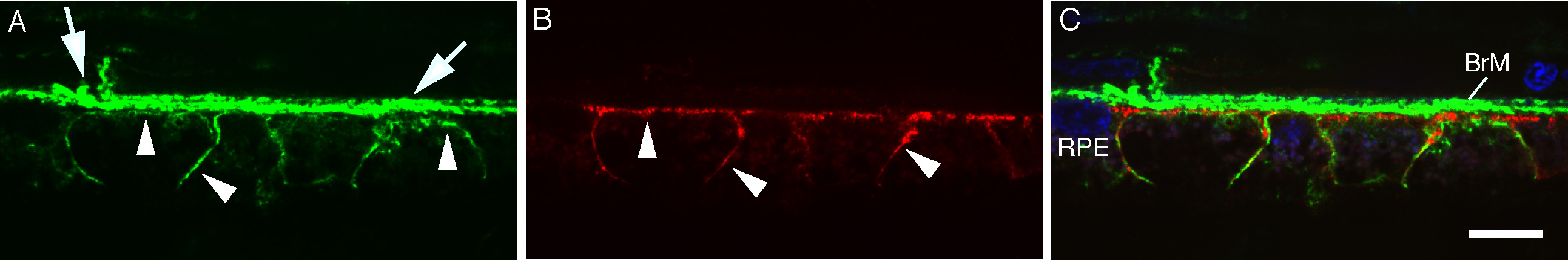
 Figure 2 of
Kochounian, Mol Vis 2016; 22:213-223.
Figure 2 of
Kochounian, Mol Vis 2016; 22:213-223.