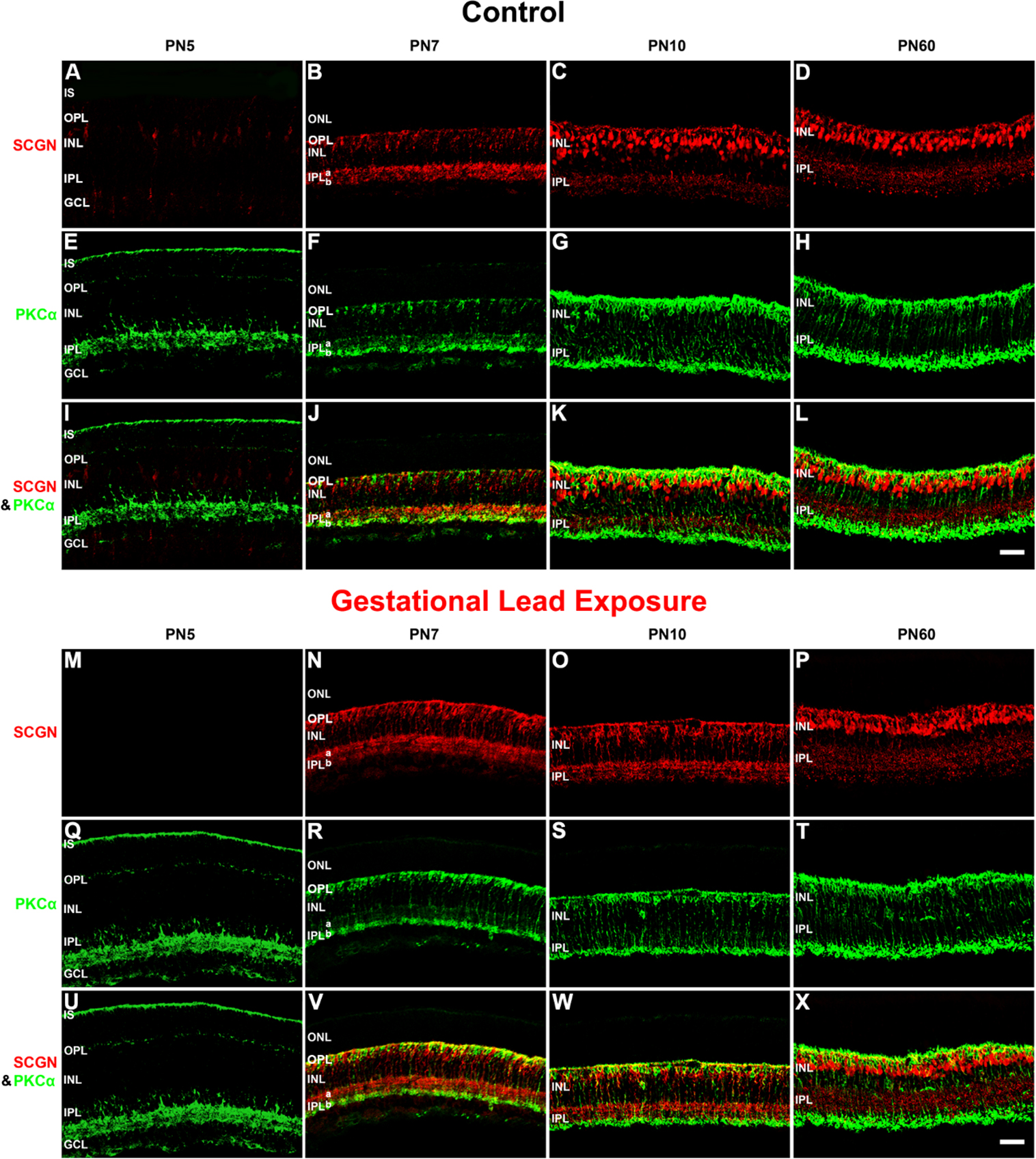
Figure 9. GLE delayed the differentiation of SCGN-IR cone BCs. The developing retinas from (
A–L) the control and (
M–X) GLE mice were double labeled with antibodies against SCGN (red:
A–D and
M–P) and PKCα (green:
E–H and
Q–T), and colabeling was examined in the merged images (yellow:
I–L and
U–X).
A-D: In the PN5 controls, the SCGN labeled cone BC somas in the INL, their dendritic processes in the OPL, and processes in the
GCL. At PN7, SCGN-IR increased and localized to immature cone BC somas in the INL, dendritic processes in the OPL, and axonal
terminals in the OFF layer (IPL-a) and the ON layer (IPL-b). SCGN-IR was more intense in IPL-a than in IPL-b. At PN10, SCGN
strongly labeled cone BC somas located throughout the INL, dendrites, and axon terminals. By PN60, SCGN-IR cone BC somas were
in the distal half of the INL, and the OPL and IPL reached their adult pattern and thickness.
E–H: The pattern of PKCα-IR in the developing controls was similar to that shown in
Figure 7.
I–L: Although SCGN and PKCα were in close approximation in the proximal IPL and especially in the OPL, they did not colabel.
M–P: In the PN5 GLE retinas, there were no SCGN-IR cells or processes. At PN7, SCGN-IR dramatically increased and was localized
to immature cone BC somas in the INL, the OPL, IPL-a, and IPL-b, the former exhibiting more intense labeling. Relative to
the age-matched controls, the INL was thicker and the IPL was less organized. At PN10, SCGN moderately labeled cone BC somas
located throughout the INL, dendrites, and axon terminals. Relative to the age-matched controls, the INL and IPL were less
developed and organized (
Table 3). By PN60, SCGN-IR cone BC somas were in the distal half of the INL, and the OPL and IPL reached their adult pattern and
thickness. Relative to the age-matched controls, the OPL, INL, and IPL were significantly thicker.
Q–R: The pattern of PKCα-IR in the developing GLE retinas was similar to that shown in
Figure 7.
U–X: Although SCGN and PKCα were in close approximation in the proximal IPL, and especially in the OPL, they did not colabel.
Scale bar = 40 μm. GLE =Gestational lead exposure; SCGN = secretagogin; IR = immunoreactive; BC = bipolar cell; PN = postnatal;
INL = inner nuclear layer; OPL = outer plexiform layer; IPL = inner plexiform layer; PKCα = protein kinase c alpha.
 Figure 9 of
Chaney, Mol Vis 2016; 22:1468-1489.
Figure 9 of
Chaney, Mol Vis 2016; 22:1468-1489.