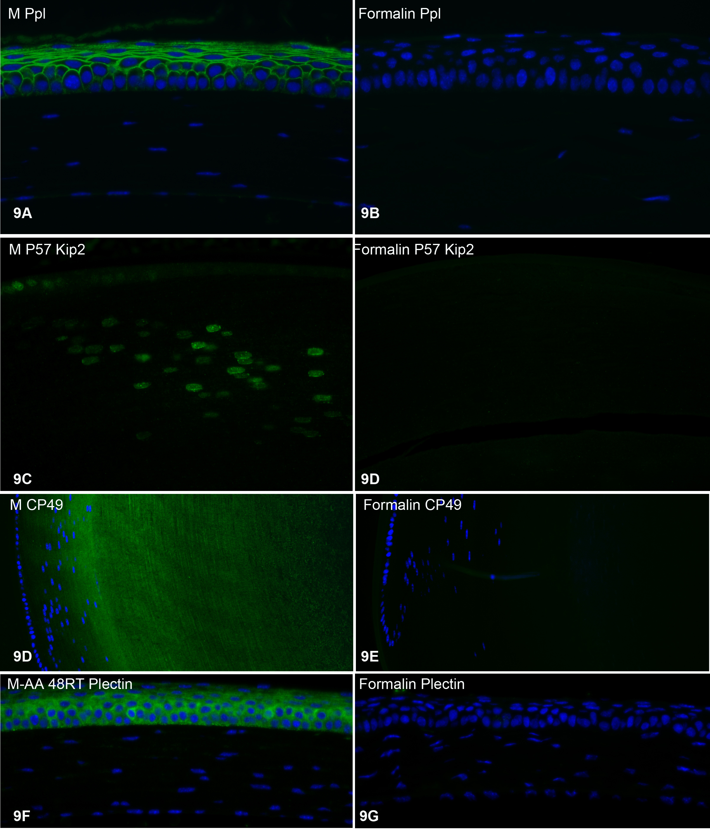
Figure 9. Labeling with four additional polyclonal antibodies.
A,
C,
D, and
F were all prepared by freeze substitution;
B,
D,
E, and
G were all prepared by immersion in formalin.
Figure 9A-B: anti-periplakin labeling of the cornea.
Figure 9C-D:anti-P57 Kip2 labeling of the lens bow region.
Figure 9D-E: anti-CP49 labeling of the lens bow region.
Figure 9F-G: anti-plectin labeling of the cornea.
 Figure 9 of
Sun, Mol Vis 2015; 21:428-442.
Figure 9 of
Sun, Mol Vis 2015; 21:428-442.