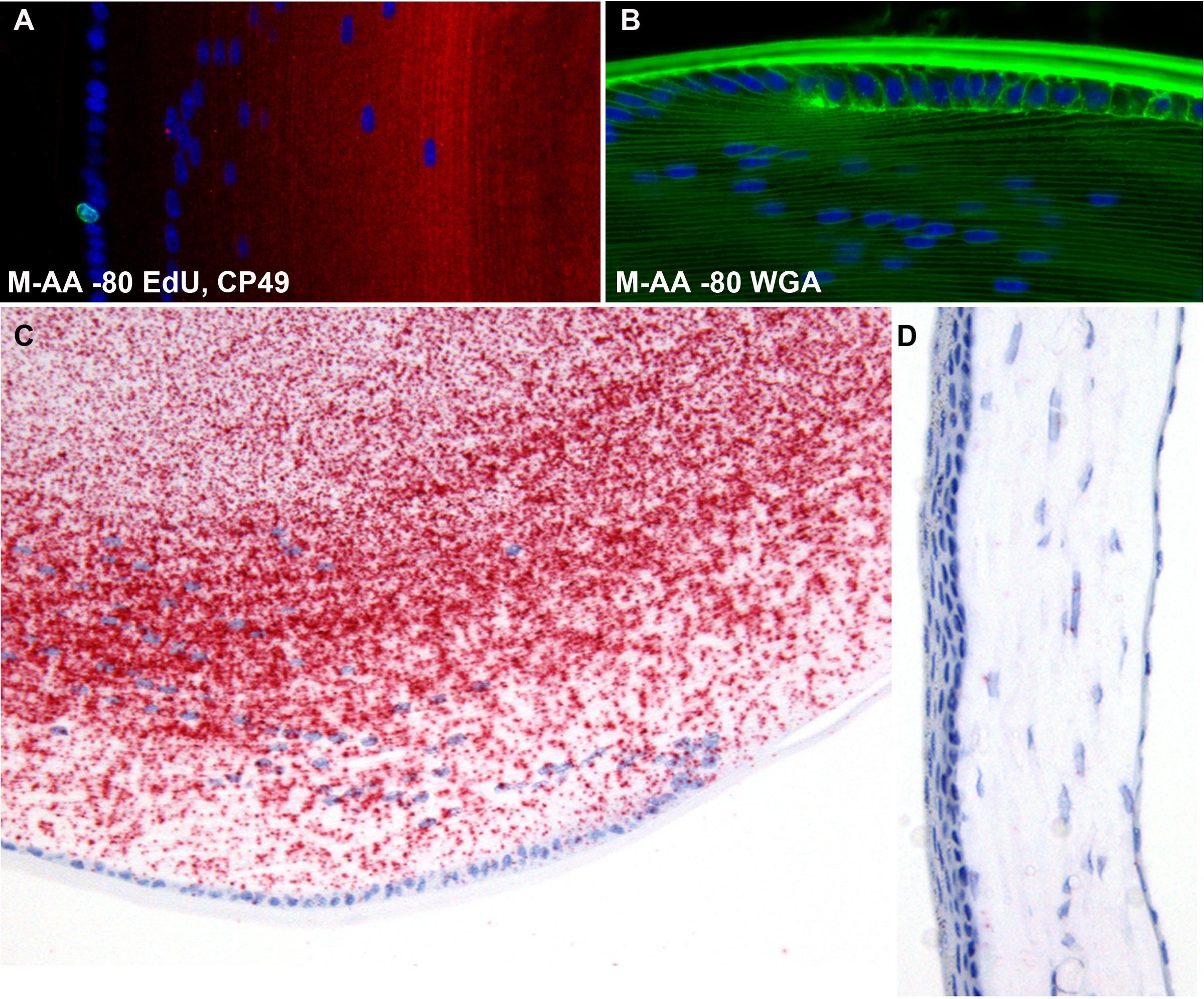
Figure 12. EdU and lectin labeling, in situ hybdridization. Panel
A demonstrates that the freeze substitution approach does not negatively affect the utility of the Click-iT chemistry approach
to the identification of dividing cells using the thymidine analog EdU. A single, Alexa fluor 488-labeled nucleus (green)
can be seen in the lens epithelium. The slide was subsequently labeled with antibodies to CP49 (red), as in
Figure 9. Panel
B shows that the labeling intensity of fluorescent lectins (WGA) is not negatively affected by the freeze substitution preservation.
To determine whether the freeze substitution methodology interfered with the capacity to conduct in situ hybridization, we
probed de-paraffinized sections from M-AA −80 samples with probes for the lens-specific CP49 mRNA. While no direct comparison
was made with formalin-fixed tissue, Panel
C clearly shows a robust signal, while the internal control from the same section (cornea, Panel
D) shows very low levels of background binding of ISH probe to CP49. The tissue processing steps required for the in situ hybridization
process introduced some loss of tissue structure, as can be seen in the slightly buckled cornea.
 Figure 12 of
Sun, Mol Vis 2015; 21:428-442.
Figure 12 of
Sun, Mol Vis 2015; 21:428-442.