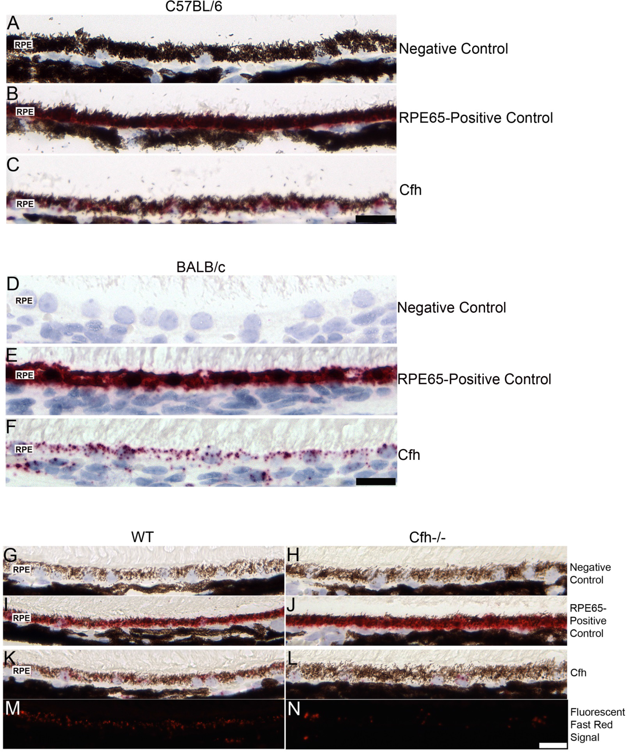
Figure 4. In situ hybridization in the C57BL/6, BALB/c, 129/Sv, and
129/Sv Cfh−/− mouse eye. A C57BL/6 eye sectioned near the optic nerve with its negative control (
A), RPE65 positive control (
B), and Cfh probe (
C) are shown. Similar in situ hybridization (ISH) results were observed in the BALB/c strain: negative control (
D), RPE65 positive control (
E), and Cfh probe (
F). Note the robust Cfh mRNA signal in the RPE in both strains (
Figure 4C,F). Negative and RPE65 positive controls and Cfh ISH results for a
129/Sv Cfh−/−mouse (
H,
J,
L) and its background strain, 129/Sv (
G,
I,
K) are shown. To visualize the Fast Red signal masked by the RPE pigment, the Fast Red fluorescence property was used to image
the sections using a rhodamine filter for 129/Sv (
M) and
129/SvCfh −/− (
N). Note the greatly reduced signal in the RPE of the
Cfh−/− eye compared to the background strain. RPE=retinal pigment epithelium. Scale bar=20 μm.
 Figure 4 of
Smit-McBride, Mol Vis 2015; 21:110-123.
Figure 4 of
Smit-McBride, Mol Vis 2015; 21:110-123.