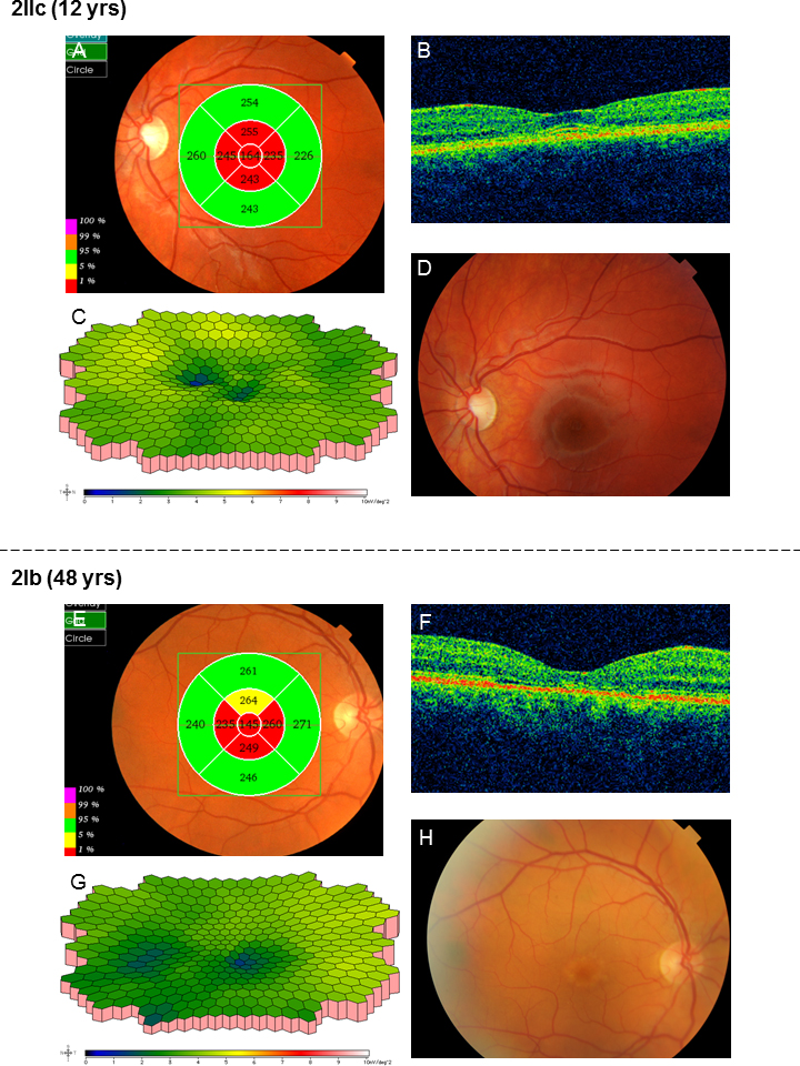
Figure 6. Results of the examinations for patients 2IIc and 2Ib in family 2.
A: In patient 2IIc, optical coherence tomography (OCT) showed attenuated retina in the innermost five segments of the macular
map even before visual acuity (VA) was reduced.
B: Moreover, intraretinal fluid was found in the center of the macula.
C: The amplitudes of the multifocal electroretinography (mERG) were reduced especially in the center compared to the normal
condition (
Figure 5I), but implicit times were normal.
D: Only subtle unspecific changes with discrete irregular macular pigmentation were found in the fundus.
E: OCT of the father in family 2, patient 2Ib, also showed attenuated retina in the five innermost segments of the macular
map although the macular contour was normal (
F).
G: The mERG showed reduced paracentral amplitudes. Implicit times were normal.
H: This patient had previously, at another clinic, been diagnosed with age-related macular degeneration due to yellow flecks
around the macula.
 Figure 6 of
Kjellström, Mol Vis 2014; 20:89-104.
Figure 6 of
Kjellström, Mol Vis 2014; 20:89-104.