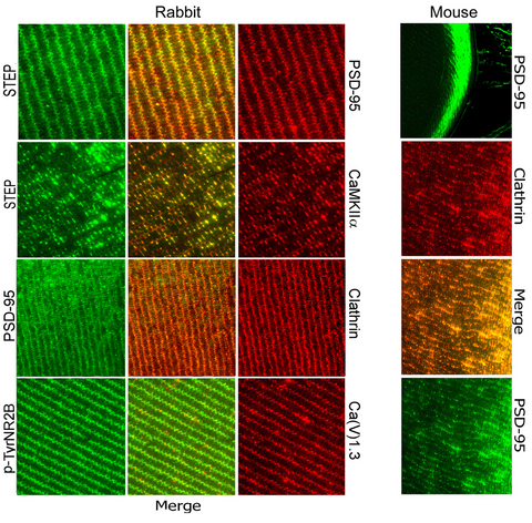
Figure 2. In situ indirect immunofluorescence detects PSD-95 in about 4-month-old rabbit and 28-day-old mouse lenses at the fiber cell
borders, overlapping with additional neuronal membrane markers shown previously to be expressed at lens fiber cell membranes.
Left panels: IF detection of PSD-95, tyrosine phosphatase STEP, CaMKIIα, clathrin, P-Tyr-NR2B, and Ca(V)1.3 in the rabbit
lens histological sections (original magnification 600X). Merged views show dual fluorescence detection demonstrated in adjacent
panels. Right panels: The top panel shows detection of PSD-95 in the mouse lens (original magnification: 200X). The three
panels below demonstrate dual fluorescence detection of PSD-95 and clathrin in the mouse lens (original magnification: 600X).
Negative controls with primary antibody are omitted since they appeared in
Figure 1.
 Figure 2 of
Frederikse, Mol Vis 2014; 20:1660-1667.
Figure 2 of
Frederikse, Mol Vis 2014; 20:1660-1667.