Figure 1. Immunolocalization of FAM18B in the human retina. A, B: Negative controls for immunohistochemistry and immunofluorescence, respectively. Autofluorescence is evident in the RPE
(white arrow). Immunohistochemistry demonstrates FAM18B (brown) in the retinal and choroidal vasculature (yellow arrows; C, E). Immunofluorescence reveals a prominent signal for the FAM18B (red) in the retinal and choroidal vasculature (yellow arrows; D, F).
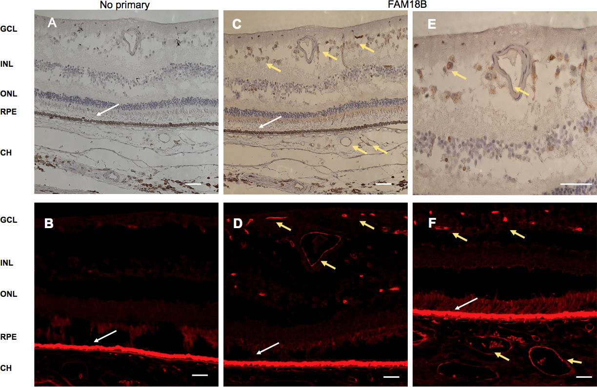
 Figure 1 of
Wang, Mol Vis 2014; 20:1146-1159.
Figure 1 of
Wang, Mol Vis 2014; 20:1146-1159.