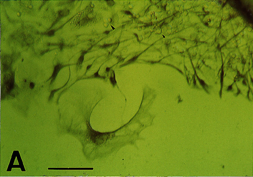
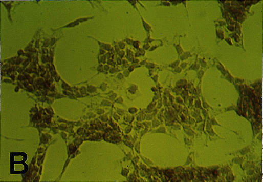
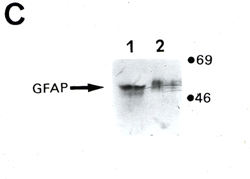
A) P6 retinal cell outgrowth from explant, 6 days in vitro . Note fibrillar staining pattern of large, flat cells and processes. Round, neuronal-like cells remain unstained (arrowheads). B) R28 cells show immunostaining to some degree throughout, in a non-fibrillar pattern. Scale Bar = 20 ”m. C) Western immunoblot for GFAP: Lane 1) P6 retina, Lane 2) R28 cells. 2 minute chemiluminescent exposure.