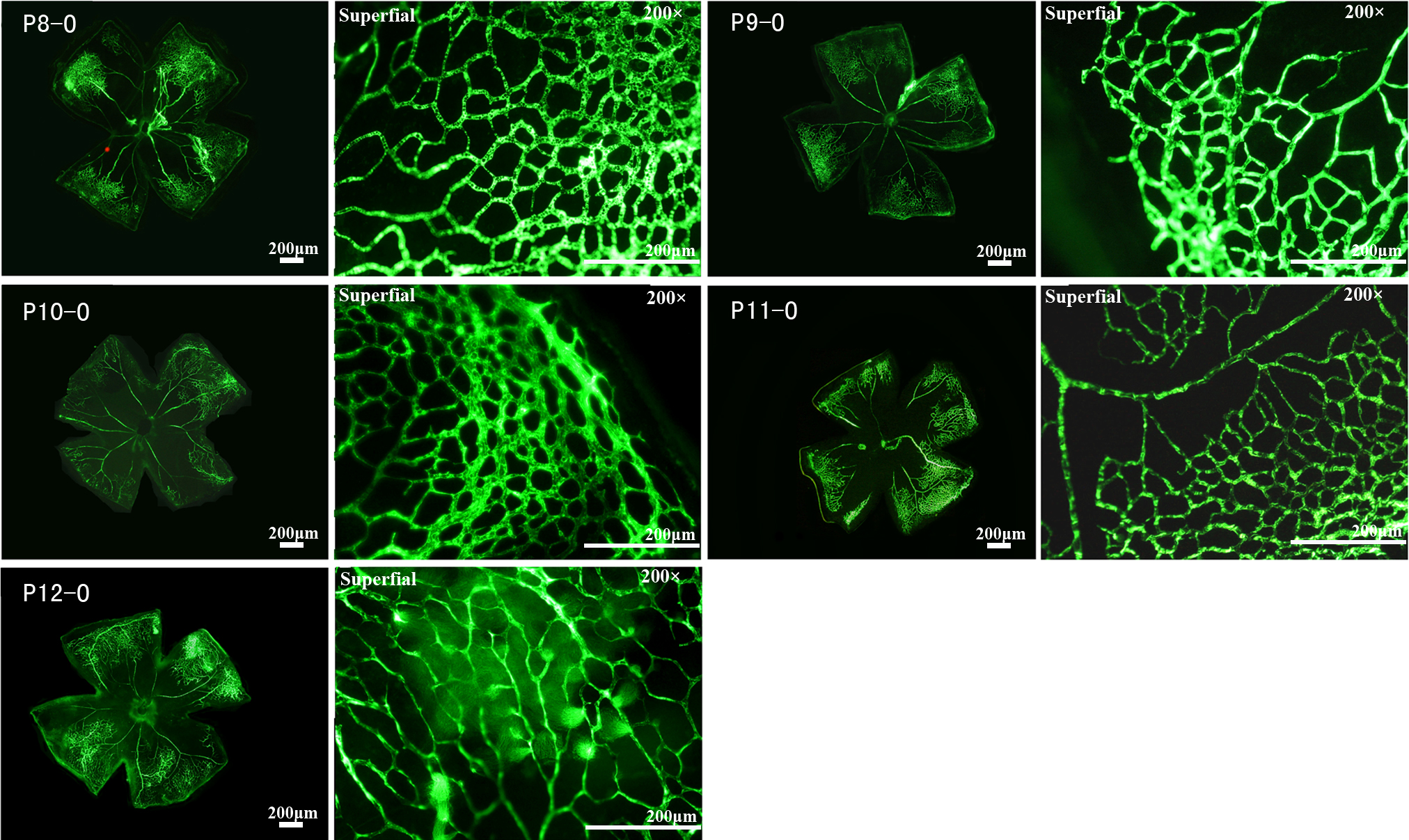
Figure 2. Central non-perfusion area and vasoconstriction under hyperoxic conditions in C57BL/6J mouse retinas. After the mice spent
24 h in a hyperoxic condition, a central area of vasoobliteration (VO) developed rapidly and increased in size until P11,
extending further into the peripheral retina, while the size of the central vasoobliteration area decreased slightly from
P12. From P8 to P12, only the superficial retinal vascular network was observed, but initiated vascular sprouts were sometimes
observed at P12. The development and differentiation of the deep and intermediate vascular plexuses were obviously suppressed.
The clear, sharp appearance of all vessels at the single superficial level in all panels also implied the lack of the two
other layers of vasculature observed in
Figure 1.
 Figure 2 of
Yang, Mol Vis 2013; 19:775-788.
Figure 2 of
Yang, Mol Vis 2013; 19:775-788.