Figure 1. Normal development of the vascular plexuses of C57BL/6J mouse retinas. Following FD-2000S perfusion, retinal whole mounts
were examined to assess the distribution of vessels along the width and depth. Great caution was needed to differentiate the
vessels at different levels; this differentiation was achieved by continuous slow adjustments of the microscope focus knob.
The specified vasculatures (superficial, intermediate, and deep) in the pictures were highlighted by tracing the vessels in
red using Adobe Photoshop software. A: Observation of retinal vessels as wholemount. From P0 to P7, only superficial retinal vessels were seen to originate from
the optic disc, extending rapidly from the inner retina to the peripheral retina during the first week of postnatal development.
B: Observation of vessels focusing on different layers of the retinal vasculature. From P8, a deep, intermediate vascular plexus
started to develop. The deep vascular plexus began forming from vertical vessels diving down from the superficial vascular
plexus, expanding from the inner retina into the peripheral retina. In addition, between P8 and P10 the intermediate vascular
plexus had not begun to form. At P12, the intermediate vascular plexus was observed, and between P15 and P17, the superficial,
intermediate, and deep vascular plexuses were seen all over the retina. After P21, the retinal vessel network was obviously
remodeled, especially the intermediate vessel layer, and no obvious changes in the retinal vessels were observed after P25.
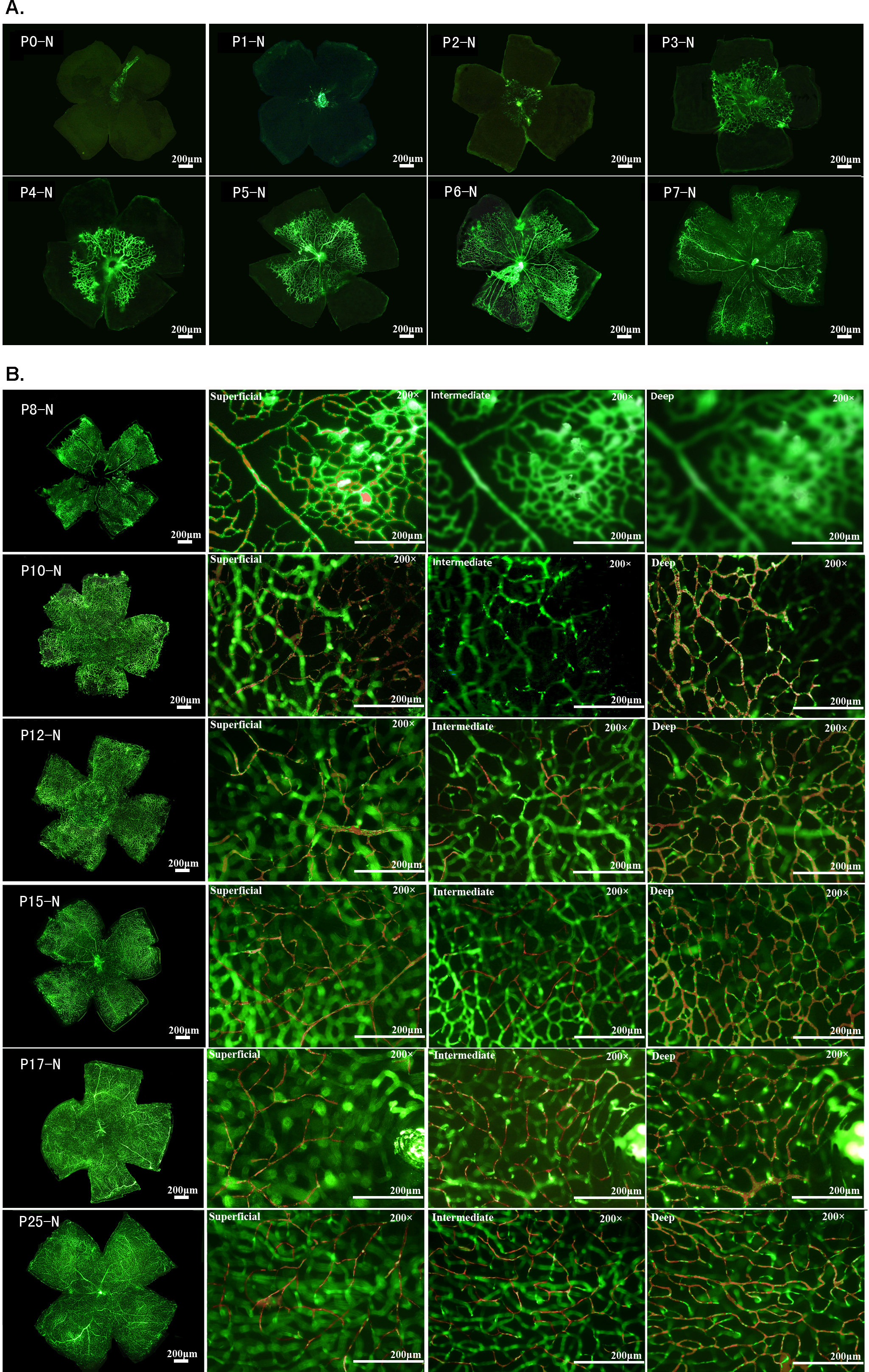
 Figure 1 of
Yang, Mol Vis 2013; 19:775-788.
Figure 1 of
Yang, Mol Vis 2013; 19:775-788.