Figure 2. Gel-filtration chromatography and gel electrophoresis were used to fractionate lens extracts and characterize different crystallin
families.
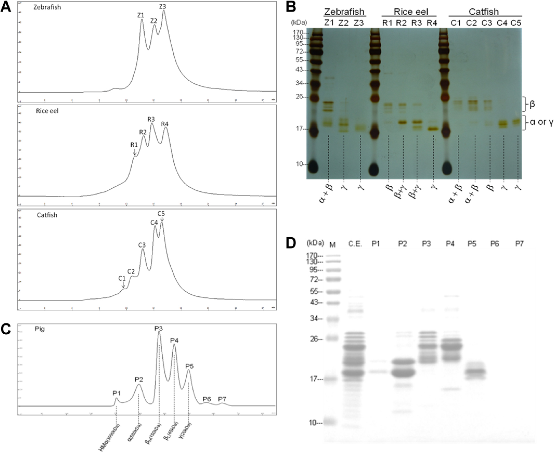
A: Comparative gel-filtration chromatography on the TSK-G4000SW
XL size-exclusion column of lens extracts from the lenses of three piscine species (two nocturnal and one diurnal). Conditions
were as described in “Materials and methods.” The column eluates (0.75 ml/tube per min) were monitored for absorbance at 280
nm. The 3–5 peaks (Z1-Z3, R1-R4, and C1-C5) in the middle of the figures correspond to the separated crystallin fractions
of three fish lenses. The absorbances at 280 nm (ordinates) shown are relative concentrations in arbitrary units.
B: Gel electrophoresis of the isolated crystallin fractions under denaturing conditions in the presence of 5 mM dithiothreitol
(sodium dodecyl sulfate–polyacrylamide gel electrophoresis [SDS–PAGE]). Lanes Z1-Z3, R1-R4, and C1-C5 correspond to the three
crystallin fractions of zebrafish, four crystallin fractions of rice eel, and five crystallin fractions of catfish in
Figure 2A. The gels were stained with silver stain. The enclosed regions denoted by α, β, or γ on the right side indicate the subunit
positions for α- and γ-crystallins with a molecular mass of about 20 kDa and β with molecular masses in a range of 24–32 kDa.
The values in kDa for the far-left lane indicate the positions of protein markers with known molecular masses.
C: Gel-filtration chromatography on the TSK-G4000SW
XL size-exclusion column of lens extracts from porcine lenses. The elution conditions are the same as those in
Figure 2A. P1-P5 fractions correspond to the five mammalian crystallin fractions of HMα, α, βH, βL, and γ crystallins, respectively
[
32]. P6 and P7 are nonprotein small molecules.
D: The isolated crystallin fractions in
Figure 2C were analyzed by gel electrophoresis under denaturing conditions in the presence of 5 mM dithiothreitol (SDS–PAGE). P1-P5
correspond to five crystallin fractions and crude extract (CE) of porcine lenses shown in
C. The gels were stained with Coomassie blue G-250.
 Figure 2 of
Lin, Mol Vis 2013; 19:623-637.
Figure 2 of
Lin, Mol Vis 2013; 19:623-637.