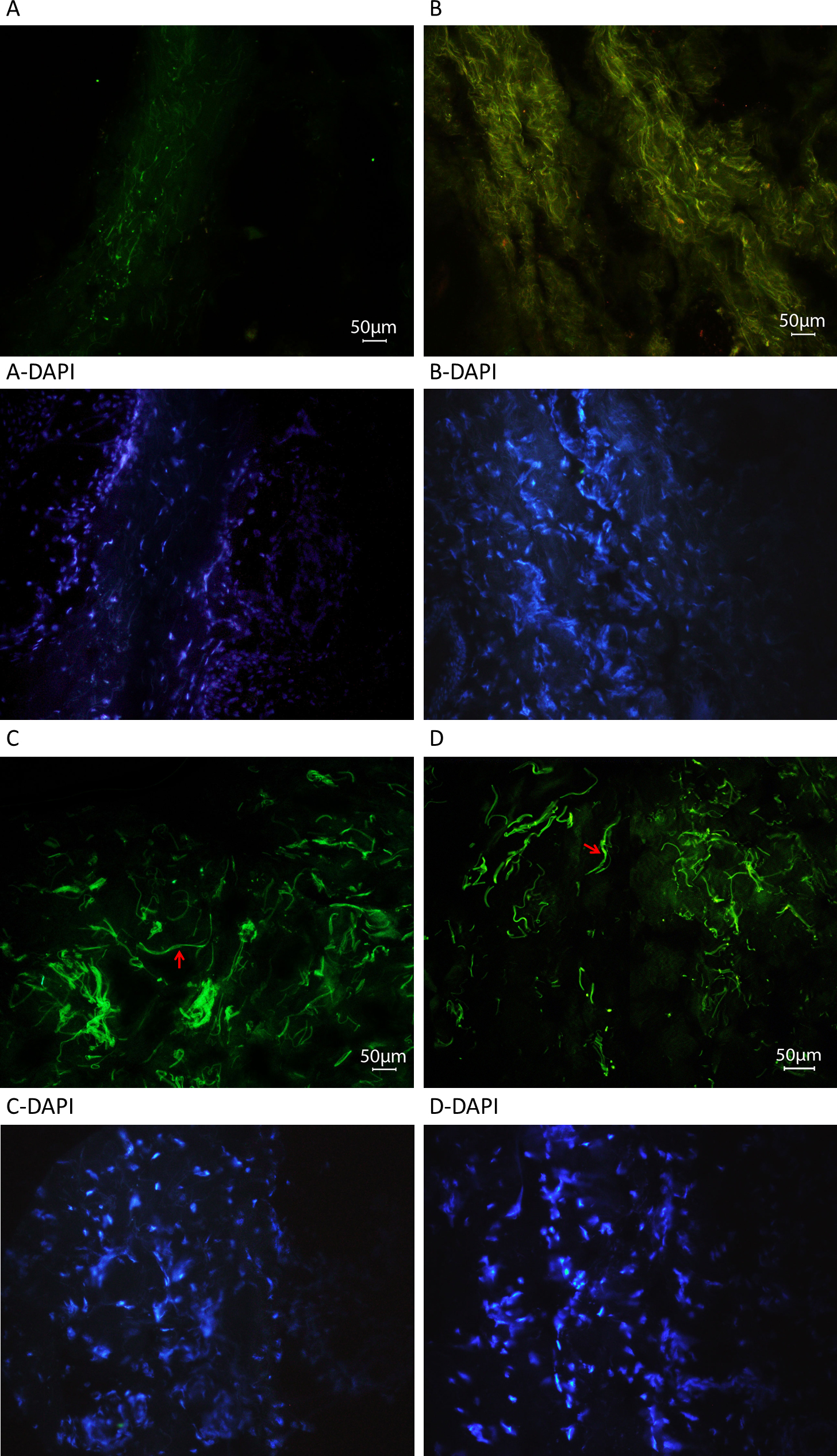
Figure 8. Fluorescent microscope images of skin tissue sections from patients with p.Tyr1792fsX55-causing mutations and age-matched
control individuals stained for latent transforming growth factor-beta binding protein 2. Representative immunofluorescent
cryosections from a homozygous carrier with primary congenital glaucoma (PCG;
A) and the control individual (
C), and from a heterozygous carrier with pseudoexfoliation (PEX) syndrome (
B) and the control individual (
D). Fibers stained for latent transforming growth factor-beta binding protein 2 (LTBP2) in the patient with PCG are thinner
and noticeably fewer (
A) compared with the control individual (
C). Fibers in the patient with PEX syndrome are also thinner, but dense and convoluted (
B) compared to the control individual (
D). Examples of longer thicker fibers in the control sections are shown with arrows; these were not seen in the sections of
patients’ tissues. Negative control is shown in
Figure 9.
 Figure 8 of
Jelodari-Mamaghani, Mol Vis 2013; 19:333-347.
Figure 8 of
Jelodari-Mamaghani, Mol Vis 2013; 19:333-347.