Figure 9. Fluorescent microscope images of skin tissue sections from patients with p.Tyr1792fsX55-causing mutations and age-matched
control individuals stained for fibrillin-1. Representative immunofluorescent cryosections from a homozygous carrier with
primary congenital glaucoma (PCG; A) and the control individual (C), and a heterozygous carrier with pseudoexfoliation (PEX) syndrome (B) and the control individual (D). The thick and long fibers that stained for fibrillin-1 in the control individuals (C, D; arrows) were not observed in multiple sections derived from the patient tissues (A, B). Fibers in the patient with PEX syndrome are dense and convoluted (B) compared to the control individuals (D). Negative control is shown in E.
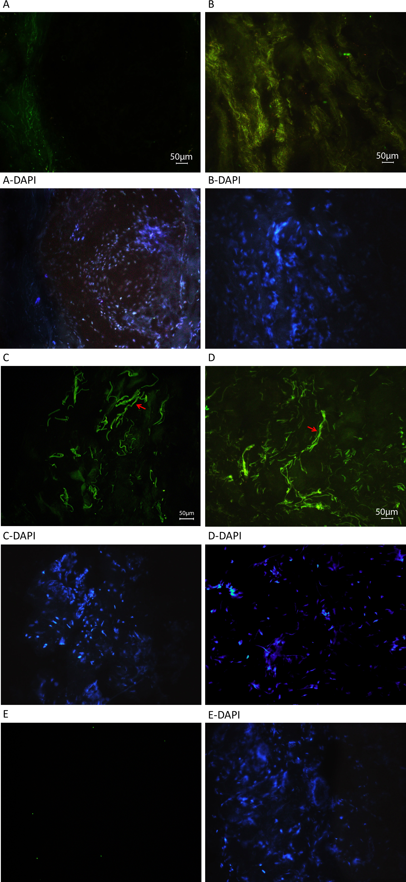
 Figure 9 of
Jelodari-Mamaghani, Mol Vis 2013; 19:333-347.
Figure 9 of
Jelodari-Mamaghani, Mol Vis 2013; 19:333-347.