Figure 2. Composite fundus photograph showing several aspects of the avascular vitreous veil (arrows and white box), a hallmark feature
of Wagner syndrome. A: The color fundus photograph of the proband’s right eye (individual II:1) shows a well-defined circumferential veil. B: Red-free fundus photograph of the proband’s left eye (Individual II:1) shows the vitreous veil as well as bright reflective
areas corresponding to peripheral chorioretinal atrophy. C: Photograph of the proband’s mother left eye (individual I:2) also shows a barely detectable veil. D: Enlargement of the area within the box shows the veil firmly attached to the underlying retina. E: Photograph of the proband’s sister right eye (individual II:5) shows a barely detectable veil more easily seen in the inferotemporal
retinal periphery.
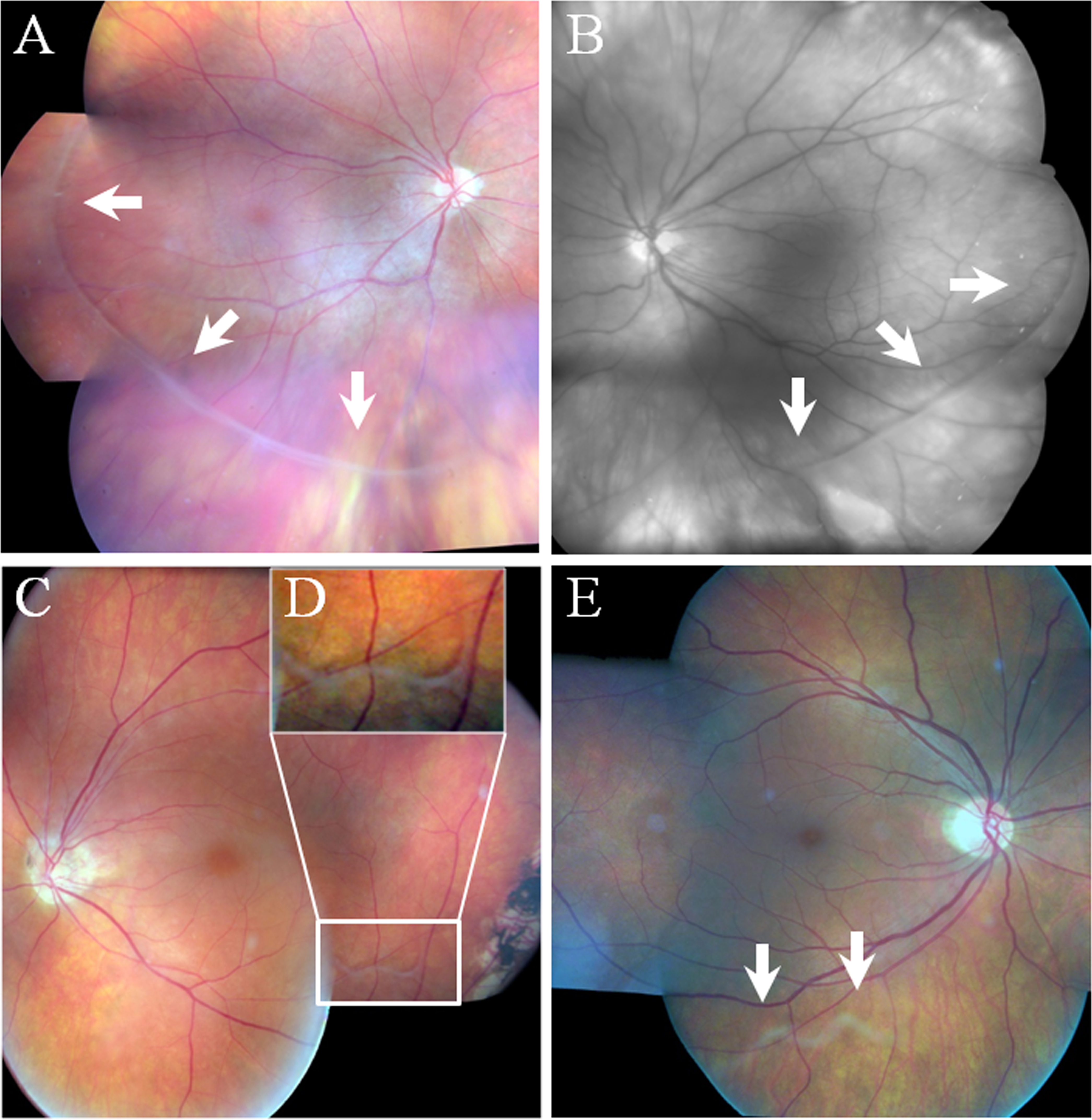
 Figure 2 of
Rothschild, Mol Vis 2013; 19:2040-2049.
Figure 2 of
Rothschild, Mol Vis 2013; 19:2040-2049.