Figure 1. Ophthalmic and genetic analysis of a family with Wagner syndrome. A: Anterior segment photograph of the proband’s right eye (individual II:1) showing the synechiae between the intraocular lens
and the iris, a temporally subluxated intraocular lens (IOL) as well as anterior and posterior capsular fibrosis. B: Pedigree of the family with Wagner syndrome. The arrow designates the proband, and the asterisks point to the family members
examined and sequenced at our center. The symbol “M/+” designates patients with the mutation. C: Normal sequence chromatogram of the versican (VCAN) gene. D: Forward and reverse mutated sequence chromatograms (individual II:1) of the VCAN gene shows a heterozygous T to A substitution (red arrow with the letter “M”) at the sixth base of the splice acceptor site
of intron 7 (c.4004–6T>A). Note that for all patients the mutated forward sequence was of poorer quality that of the reverse
sequence.
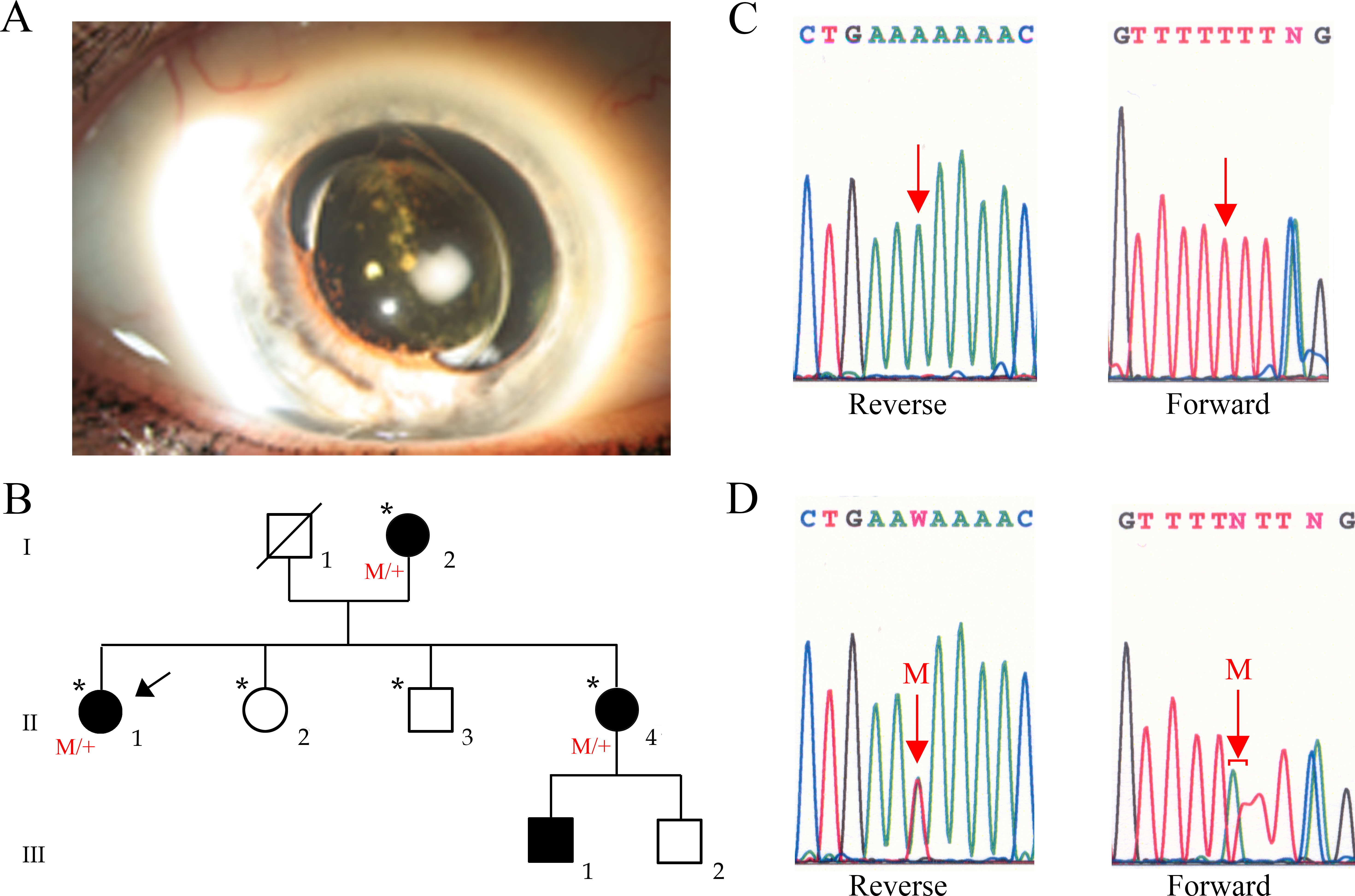
 Figure 1 of
Rothschild, Mol Vis 2013; 19:2040-2049.
Figure 1 of
Rothschild, Mol Vis 2013; 19:2040-2049.