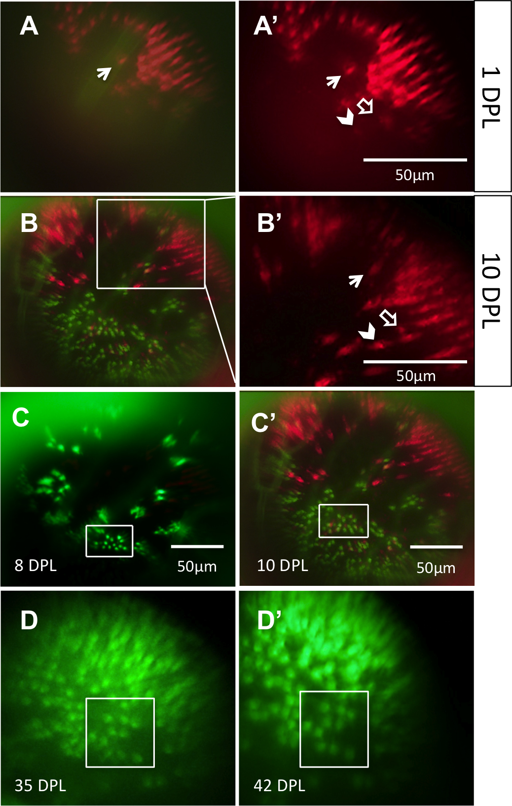
Figure 4. The same locations on the retina can be recovered and viewed on subsequent days. Through a combination of approximate relocation
using vessel branch patterns and more precise relocation using unique lesion edge patterns, areas of interest were reexamined
at multiple time points.
A: In the mCherry channel (blue-sensitive cones), a lesion edge reminiscent of a bay shape, and a single cone within the bay
(arrow), was observed at 1 day post-lesion (DPL). Image in A is a merge of the green fluorescent protein (GFP) and mCherry
channels;
A’ shows the mCherry channel alone.
B: The same bay-shaped lesion edge was relocated at 10 DPL.
B is a merge of the GFP and mCherry channels; B’ shows a higher magnification of the white box in
B, in the mCherry channel alone. The single cone (arrow) was also identified. The appearance of a new blue-sensitive cone between
1 DPL [
A’] and 10 DPL [
B’] is indicated with an empty arrow. The chevron indicates a potential surviving cone, but the faint signal from this location
at 1 DPL puts to question if the cone died and was replaced by the bright cone seen at 10 DPL.
C: An area containing uniquely-shaped patches of surviving ultraviolet-sensitive cones is observed at 8 DPL (top) and relocated
at 10 DPL (bottom). The same collection of ultraviolet-sensitive cones, and the same individual cones, was identified (white
boxes) at both days.
D: An area of regenerated UV cones showing the loss of row mosaic organization, imaged at 35 and 42 DPL (panel
D and
D’, respectively); matching clusters of cones are indicated with white boxes. For an example of the outlined vessels and how
areas were located using vasculature, see
Figure 3. n=2 fish shown.
 Figure 4 of
Duval, Mol Vis 2013; 19:1082-1095.
Figure 4 of
Duval, Mol Vis 2013; 19:1082-1095.