Figure 3. Using vasculature patterns as in vivo landmarks to navigate within the eye. A: An in vivo view of the optic nerve head (ONH) is represented by the apparent convergence of vessels. From the ONH, vessel
branching patterns were utilized to record and relocate areas of interest. B: A low-magnification image of the green fluorescent protein (GFP) channel is presented, showing the branching pattern of
vessels that is used to navigate during imaging. Due to the difference in focal planes of the vessels and photoreceptor layer,
the apparent positions of individual photoreceptors may shift slightly relative to the vessels. C: The same image in B is presented with GFP and mCherry channels merged and vessels outlined in white for clarity [C’]. Due to discrepancies in focal plane between the photoreceptor layer and the vessels, relative positions of some small
areas of interest and neighbouring vessels differ slightly between images and between magnifications. To illustrate this,
an area of interest, a bay-shaped lesion edge, was imaged at different magnifications. In C’, the area of interest is located between two vessel branches (white box). D: The area, imaged at high magnification, is shown without [D] and with vessels outlined [D’]. At high magnification, the area lies underneath one of the vessel branches (arrows indicate the same branch in each image),
an apparent a shift of approximately 57 μm. n=2 fish shown.
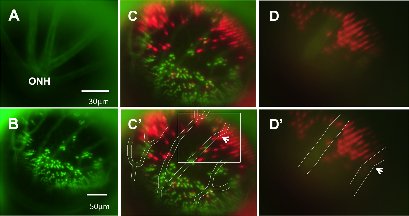
 Figure 3 of
Duval, Mol Vis 2013; 19:1082-1095.
Figure 3 of
Duval, Mol Vis 2013; 19:1082-1095.