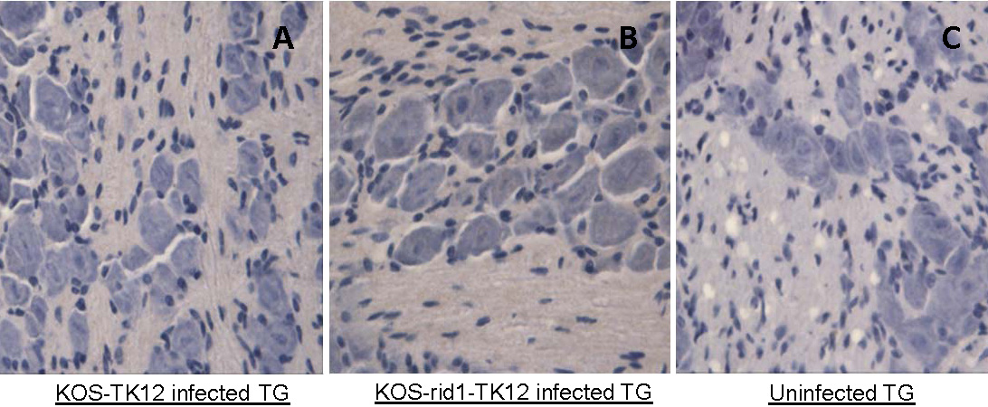
Figure 2. Expression of herpes simplex virus 1 proteins in the trigeminal ganglia of BALB/c mice. Five days after virus inoculation,
murine trigeminal ganglia (TG) were isolated, formalin fixed, paraffin embedded, sectioned and stained with a rabbit antiserum
raised against herpes simplex virus 1 (HSV-1). A lack of brown staining indicates lack of HSV-1 proteins. TG tissue 5 days
after KOS-tk12 inoculation is shown in panel A and TG 5 days after KOS-Rid1-tk12 inoculation is shown in panel
B Uninfected TG section is shown in panel
C HSV-1 infected corneas were used as positive control (
Figure 1). Representative pictures are shown.
 Figure 2 of
Shukla, Mol Vis 2012; 18:2711-2716.
Figure 2 of
Shukla, Mol Vis 2012; 18:2711-2716.