Figure 1. Histopathological changes and the expression of herpes simplex virus 1 proteins in the corneas of BALB/c mice. Formalin-fixed,
paraffin-embedded, murine ocular tissues were sectioned and stained with a rabbit antiserum raised against herpes simplex
virus 1 (HSV-1). The brown staining indicates HSV-1 proteins. Corneas 2 days after virus inoculation are shown in panel (A) KOS-tk12 and panel (B) KOS-Rid1-tk12. Uninfected cornea section is shown in panel (C) Representative pictures are shown.
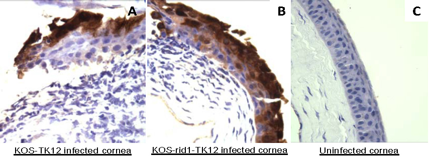
 Figure 1 of
Shukla, Mol Vis 2012; 18:2711-2716.
Figure 1 of
Shukla, Mol Vis 2012; 18:2711-2716.