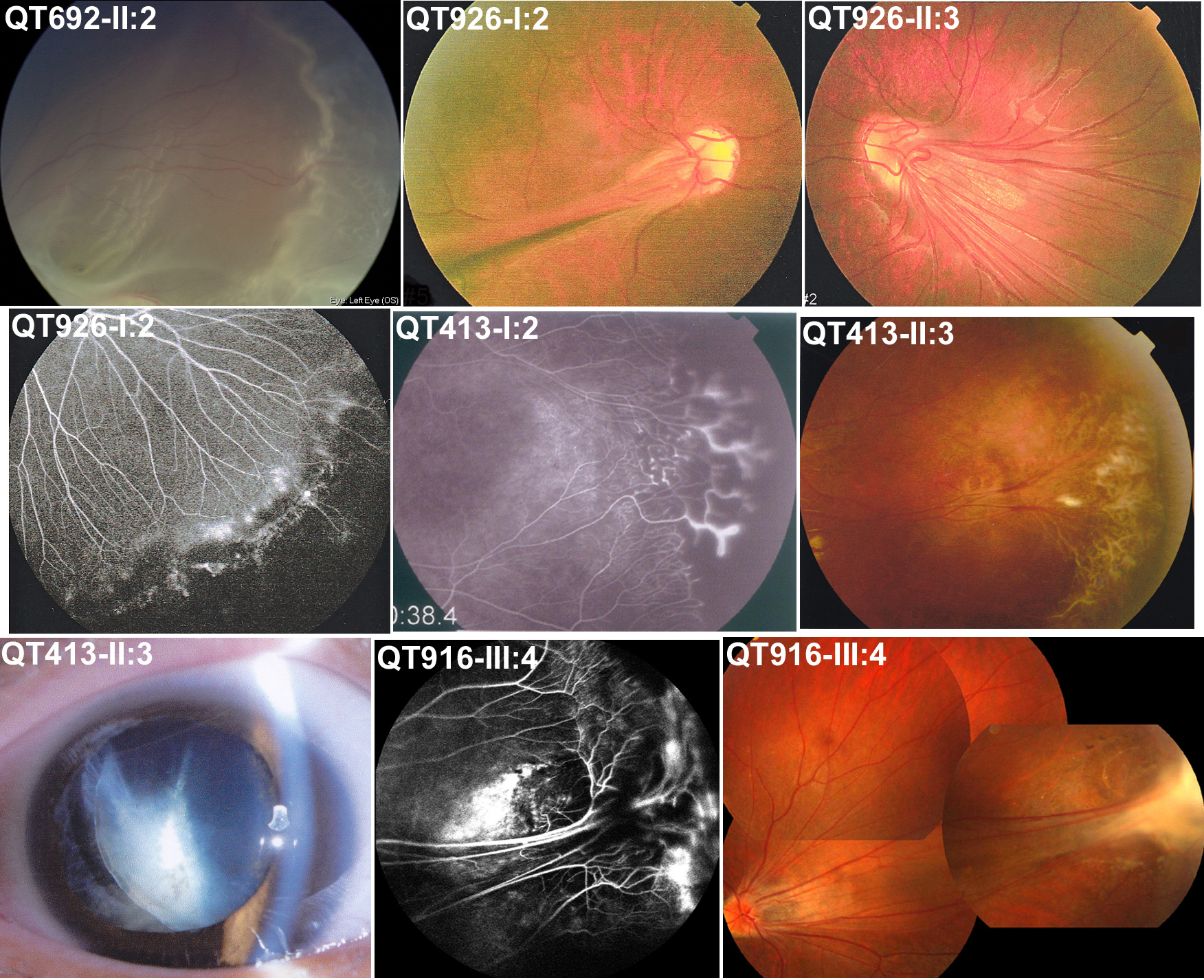
Figure 4. Ocular changes in affected
individuals with an
FZD4 or
LRP5 mutation. The
individual ID is indicated on the top left of each picture,
which is the same as in
Figure 2 and
Table 2.
Signs of FEVR included retinal detachment (top left), falciform
retinal fold (top middle), temporal dragging of optic disc (top
right), peripheral avascular zone and brush-like peripheral
vessels (middle left), shell-like peripheral vessel terminatio
and neovascularization (center), peripheral fibrovascular
proliferation (middle right), lens dislocation (bottom left),
peripheral exudates (bottom middle), temporal dragging of optic
disc, and peripheral fibrous proliferation (bottom right).
 Figure 4
of Yang, Mol Vis 2012; 18:2438-2446.
Figure 4
of Yang, Mol Vis 2012; 18:2438-2446.